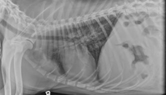
This 1-year-old dog has a history of chronic vomiting which worsened recently. What are your findings? … [Read more...]
13 year old Staffordshire Terrier
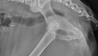
This week's case is a 13-year-old Staffordshire Terrier with a palpable pelvic mass. … [Read more...]
2 year old Thoroughbred
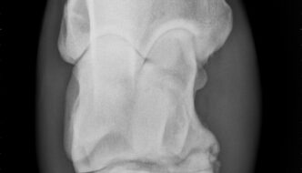
This week's imaging case comes from large animal radiology. It's a 2-year-old Thoroughbred filly that was kicked on the left tarsus. Post your interpretations in the comments section! … [Read more...]
3 year old Domestic Longhair
This week's case is a 3-year-old domestic long haired cat with suspected trauma. Post your interpretations in the comments section. … [Read more...]
13 year old Domestic Shorthair
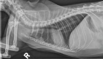
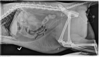
This week's case is a 13-year-old female neutered Domestic Shorthair cat with anorexia and intermittent diarrhea. Where is the primary disease located? … [Read more...]
- 1
- 2
- 3
- …
- 88
- Next Page »
Recent Comments