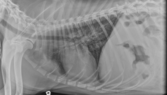
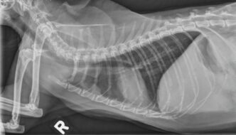
This 1-year-old dog has a history of chronic vomiting which worsened recently. What are your findings? … [Read More...]
Teaching and learning about veterinary diagnostic imaging.
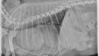
This 1-year-old dog has a history of chronic vomiting which worsened recently. What are your findings? … [Read More...]
Recent Comments