This 1-year-old dog has a history of chronic vomiting which worsened recently. What are your findings?
Findings
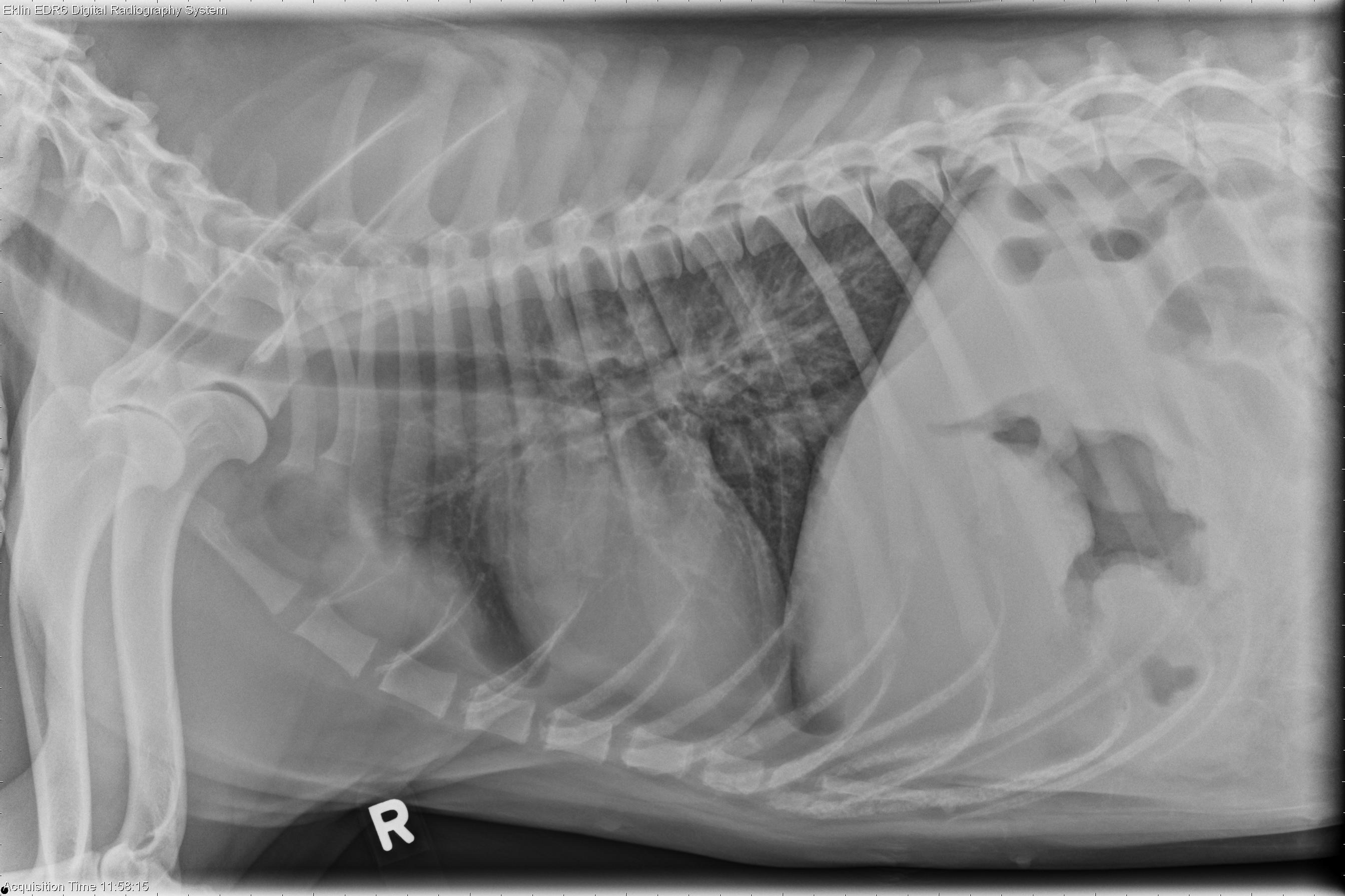
There is a convex, broad-based soft tissue opacity dorsal to the second and third sternebrae. There is a mild bronchointerstitial pattern throughout the lungs. The pleural space and musculoskeletal structures are normal. Within the abdomen, the stomach wall has an irregular contour with the suggestion of multifocal thickening. There is poor peritoneal detail with a mottled appearance. The cardiovascular structures are normal.Differential Diagnosis
- Sternal lymphadenopathy secondary to abdominal disease (reactive, neopastic)
- Probable gastric masses (neoplasia, granulomatous)
- Peritoneal effusion (transudate, exudate, hemorrhage, neoplasia)
- Bronchointerstitial pattern is nonspecific (infectious or noninfectious inflammatory, neoplastic)
Diagnosis
- Large cell granular T-cell lymphoma
- On ultrasound examination, multiple masses were identified in the gastric wall, kidney, spleen, and ureter with moderate peritoneal effusion. Necropsy revealed disseminated T-cell lymphoma. The gastric lesions were associated with ulceration, likely causing the vomiting.