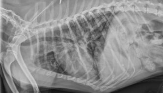
This week's case is a 6-year-old male neutered Chihuahua with chronic cough that has not responded to therapy. What are your radiographic findings? … [Read more...]
Thorax
7 year old Domestic Shorthair
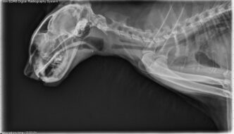
This week's case is a 7-year-old male neutered Domestic Shorthair cat with difficulty breathing. What are the two main findings? … [Read more...]
2 year old DSH
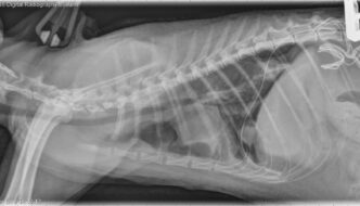
This week's case is a 2-year-old female neutered DSH cat. Missing for two days, returned depressed, dehydrated, and anorexic. What are your findings and diagnostic plan? … [Read more...]
7 year old mixed breed dog
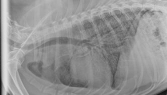
This week's case is a 7-year-old male neutered mixed breed dog with lethargy, fever, lameness, and cutaneous lesions. How would you characterize this pattern? … [Read more...]
11 year old Beagle
This week's case is an 11-year-old Beagle with right thoracic limb lameness. How would you describe this lesion? … [Read more...]
- « Previous Page
- 1
- 2
- 3
- 4
- …
- 43
- Next Page »
Recent Comments