• Home
  • About
  • Articles
  • Books
  • Contact

Veterinary Radiology

Teaching and learning about veterinary diagnostic imaging.

  • Modality
    • Radiographs
    • Ultrasound
    • CT/MRI
  • Region
    • Thorax
    • Abdomen
    • Musculoskeletal
    • Neurologic
  • Species
    • Canine
    • Feline
    • Equine
    • Exotic

Infarction of the spleen and liver: a similar ultrasonographic pattern

March 13, 2007 By Allison Zwingenberger

Infarction of the spleen or liver is a fairly rare event. But if the blood supply is reduced, a region, or the entire organ, can become infarcted. Infarction is necrosis resulting from a reduced blood supply. The main causes of infarction of the liver and spleen are malposition (torsion) and … [Read more...]

Filed Under: Ultrasound Tuesday

Interpreting ultrasound videos

March 8, 2007 By Allison Zwingenberger

Tonight we looked at 2-3 minute ultrasound videos as preparation for the board exams. No matter how many ultrasounds you've done, it's disconcerting to watch a partial scan that someone else has performed. They will be scanning with a different speed and pattern than you would, and the image may not … [Read more...]

Filed Under: Known Case Conference, Residents

Journal Club 3/7/07

March 7, 2007 By Allison Zwingenberger

Mantis P, Baines E. Computed tomography: Why use it in small animal practice? The Veterinary Journal 2007;173:237-238. Nuss K. Ultrasonography of musculoskeletal disorders in cattle: A practical tool for veterinary surgeons. The Veterinary Journal 2007;173:239-240. Ohlerth S, Scharf G. … [Read more...]

Filed Under: Journal Club

Ultrasound of linear foreign bodies

March 6, 2007 By Allison Zwingenberger

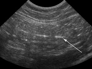
Ultrasound of linear foreign bodies can be more difficult than it seems. There are a couple of different imaging presentations depending on the size of the foreign body itself. I like to use radiographs to look for the pattern of linear foreign body, then confirm with ultrasound and look for … [Read more...]

Filed Under: Ultrasound Tuesday

Possible cranial mediastinal mass

March 1, 2007 By Allison Zwingenberger

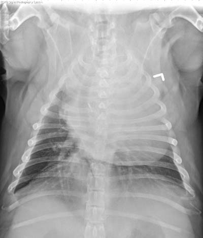
I recently had a question about diagnosing mediastinal masses. The case was a 3 year old bulldog, with an increased soft tissue opacity in the cranial mediastinum. These barrel chested dogs like bulldogs and pugs tend to store a lot of fat in the cranial mediastinum, which makes it look much wider … [Read more...]

Filed Under: Uncategorized

  • « Previous Page
  • 1
  • …
  • 177
  • 178
  • 179
  • 180
  • 181
  • …
  • 185
  • Next Page »
  • Bloglovin
  • Facebook
  • RSS
  • Twitter
AtlasCover787x1024 amazon-availablenow

Veterinary Radiology News

Sign up for alerts about new cases and newsletters.

We will respect your privacy.

Archives

Recent Comments

Tags

abscess adenocarcinoma bronchiectasis carcinoma cardiomyopathy coccidioidomycosis discospondylitis elbow dysplasia eosinophilic bronchopneumopathy erosive polyarthritis feline infectious peritonitis FIP foreign body fracture gastric dilation gastric foreign body gastrointestinal GDV heart failure hemangiosarcoma hiatal hernia histiocytic sarcoma hypertrophic cardiomyopathy intestinal foreign body linear foreign body lymphoma megaesophagus osteochondrosis osteomyelitis osteosarcoma patent ductus arteriosus PDA pericardial effusion pneumonia pneumothorax polyarthritis PPDH pulmonary pulmonary abscess pulmonary adenocarcinoma pulmonary carcinoma sarcoma thymoma tracheal collapse vascular ring anomaly

Copyright © 2025 · Metro Pro Theme On Genesis Framework · WordPress · Log in