I recently had a question about diagnosing mediastinal masses. The case was a 3 year old bulldog, with an increased soft tissue opacity in the cranial mediastinum. These barrel chested dogs like bulldogs and pugs tend to store a lot of fat in the cranial mediastinum, which makes it look much wider and more opaque than normal. But this was even more than usual. In addition, he had a markedly enlarged heart. How can you tell if there’s a mass?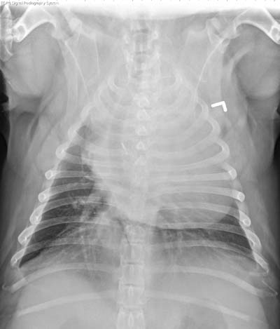
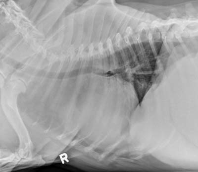
The cranial mediastinum definitely appears widened in the first set of radiographs. However, the heart is taking up the majority of the thorax, and extending into the cranial mediastinum as well. The apex of the heart is located at the left thoracic wall, meaning that the enlargement is mostly right sided. On the lateral projection, the heart is markedly enlarged, and the heart base is deviating the trachea dorsally. The carina remains in its normal position at the 5th-6th intercostal space. The trachea is also deviated to the right on the d/v projection, due to the heart size. This dog has breed-associated fat in the cranial mediastinum, as well as right heart enlargement from pulmonic stenosis.
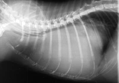
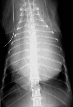
How is this different from a cranial mediastinal mass? The increased opacity in the cranial mediastinum is similar with a mass, but often appears more soft tissue dense. The dog in figure 1 has some less dense fat opacity in the mediastinum, so it’s not just uniform soft tissue opacity. Cranial mediastinal masses also displace the trachea dorsally. They tend to stay on midline, and if large enough can displace the carina caudally from its normal position. This cat has a thymoma in the cranial mediastinum which is displacing the heart caudally to the 8-9th intercostal space.
Criteria for diagnosing cranial mediastinal masses:
- Elevates trachea
- Displaces carina caudal to 5th-6th intercostal space
- Uniform soft tissue opacity in cranial mediastinum
- Midline position
- Take breed variation and other abnormalities into account