Today’s case is an 11 year old male neutered Rhodesian Ridgeback who has been choking on saliva and bringing up white foam.
Everyone has been writing great interpretations of these radiographs in the comments section, keep it up! It’s good practice to come up with findings and differential diagnoses on unknown cases.
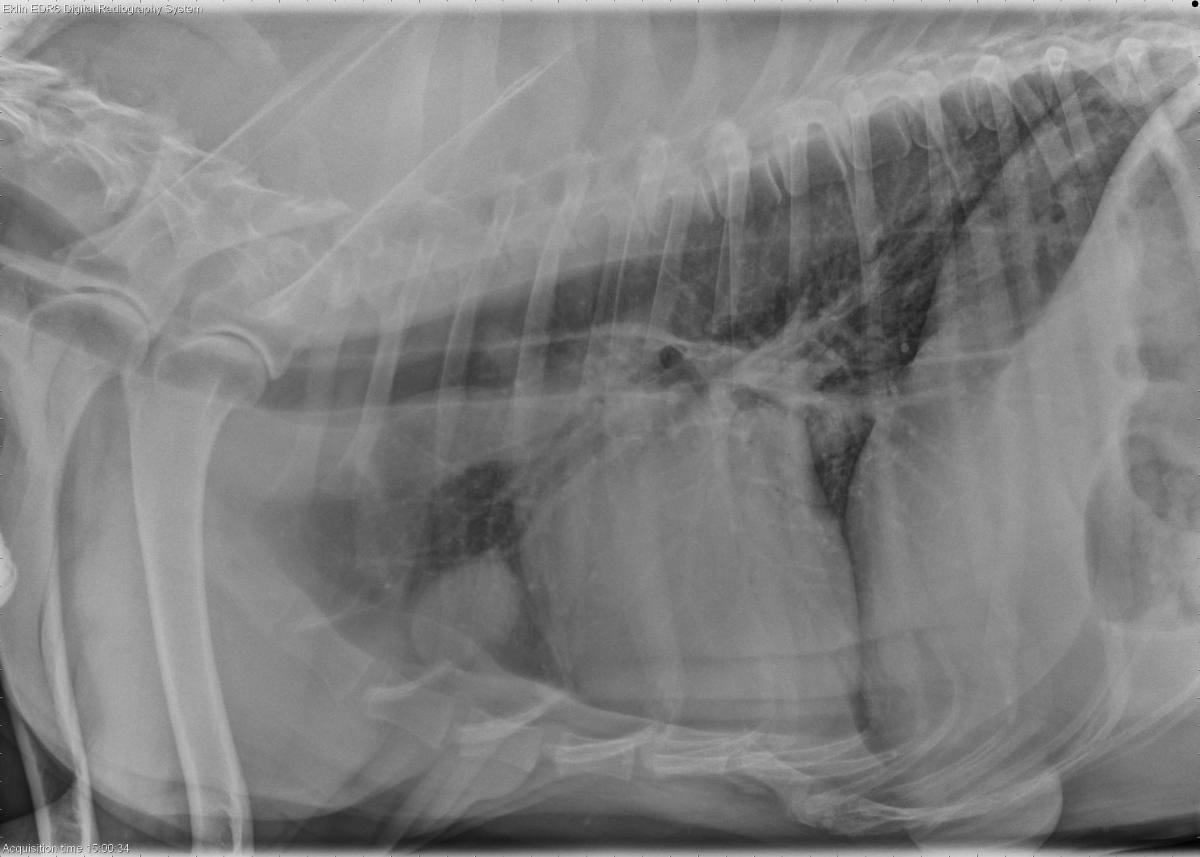
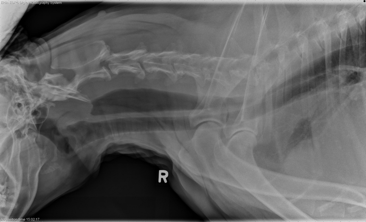
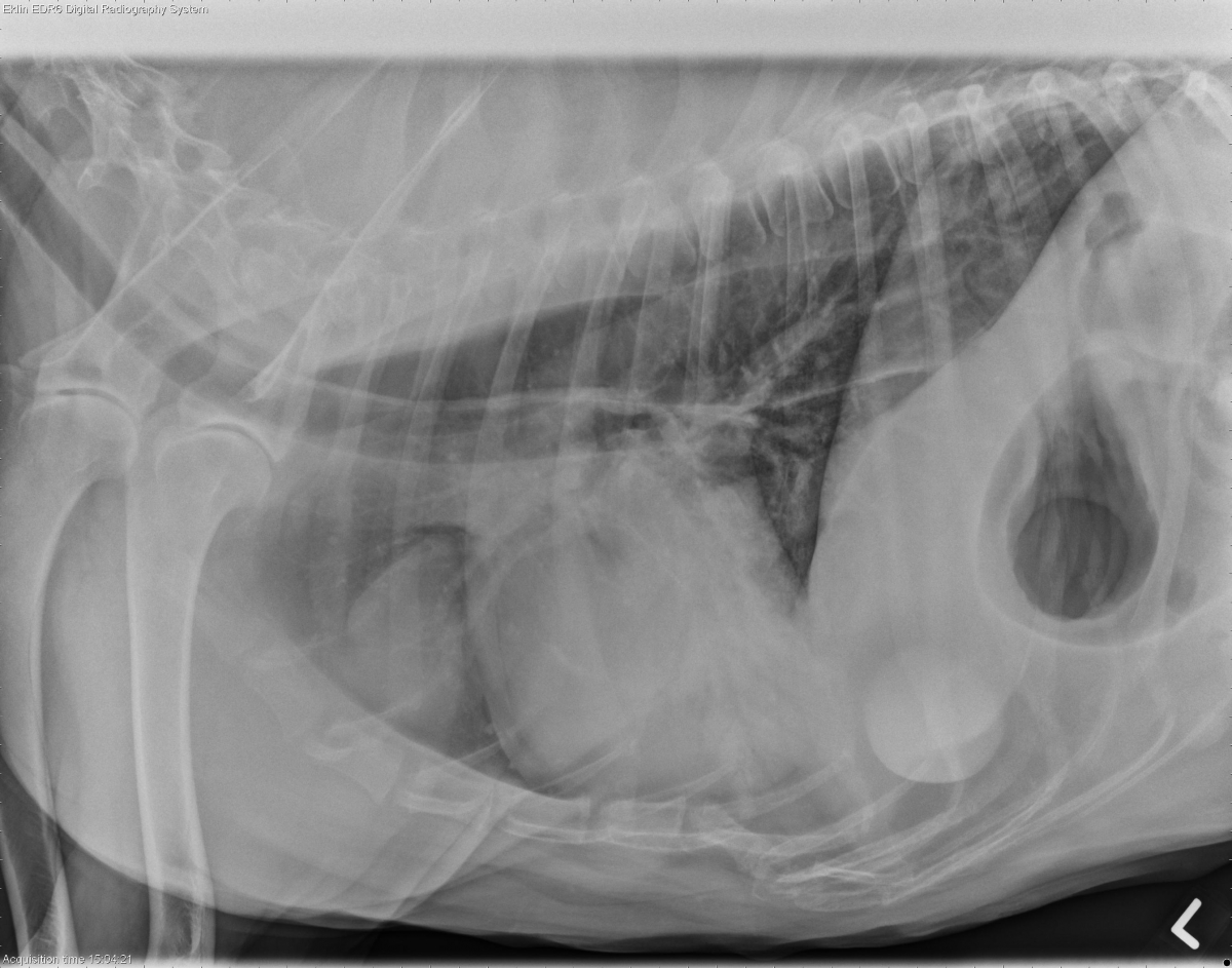
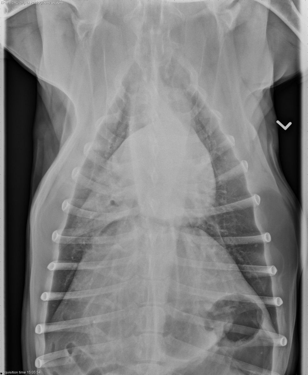
Findings
There is a rounded soft tissue mass within the cranial mediastinum just cranial to the heart. There is diffuse gas distention of the esophagus. There are alveolar infiltrates within the right middle lung lobe. There is a rounded soft tissue mass at the left ventral thoracic wall which is external to the thorax. The cardiovascular structures are within normal limits.
Differential Diagnosis
The diagnosis that ties a cranial mediastinal mass together with megaesophagus and aspiration pneumonia is a thymoma with secondary myasthenia gravis. Other causes of cranial mediastinal masses include lymph node, cyst and granuloma. Megaesophagus can be idiopathic or acquired.
Diagnosis
Thymoma with megaesophagus and aspiration pneumonia.
Discussion
Thymoma and lymphoma are the two most common causes of cranial mediastinal masses in dogs. Thymoma is associated with myasthenia gravis in 40% of dogs and occasionally in cats. Thymoma is a tumor of the epithelial portion of the thymus, but often has a population of mature lymphocytes. In contrast, thymic lymphoma is comprised of less mature lymphoblasts. On radiographs and CT, lymphoma and thymoma look similar.
- Roy ME, Wrigley R, Craft SL, et al. Radiographic prediction of the nature of cranial mediastinal masses in dogs. Journal of Small Animal Practice 2006;47:761-761.
Megaesophagus and a cranial toraxic mass
The esophagus is diffusely distended with gas. The right middle lung lobe is opaque. On the lateral views there is a soft tissue opacity cranial to the heart.
Looks like megaesophagus with aspiration pneumonia.
So the votes so far favor three major abnormalities that both readers comment on above. Where exactly is the mass cranial to the heart?
The location is suggestive of thymoma, which can also be the cause of the megaesophagus
The three abnormalities within the thorax were the megaesophagus, aspiration pneumonia, and cranial mediastinal mass. There is also a large mass on the outside of the body wall that some of you might have seen. And it’s always good to try to tie all of the abnormalities together to make one story.
Fantastic case! Any info on the mass outside the thorax? Maybe lipoma?