Today’s case is an 8-year-old male neutered Labrador Retriever with cough for 6 weeks, transient improvement with furosemide. What is your radiographic diagnosis?
Findings
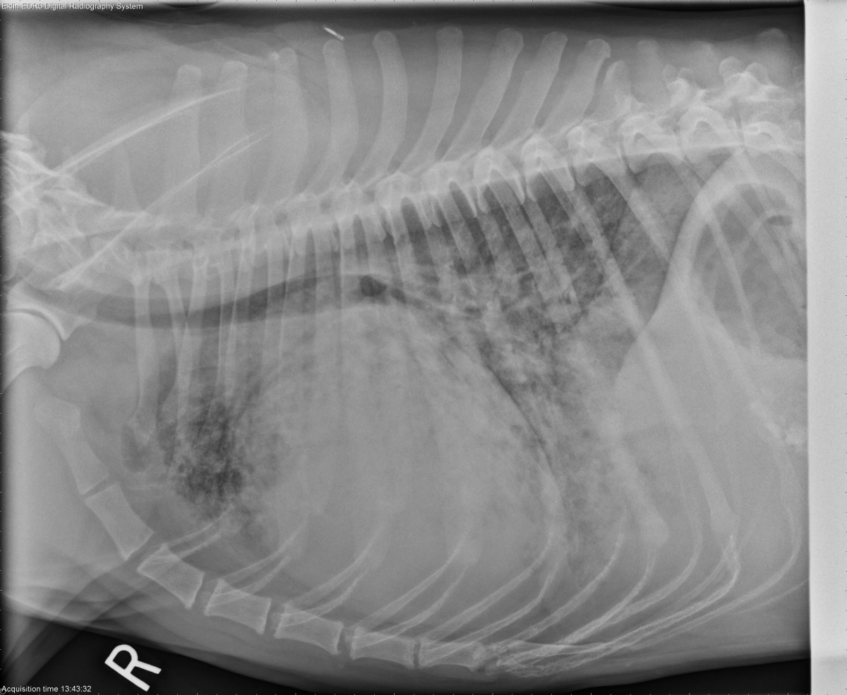
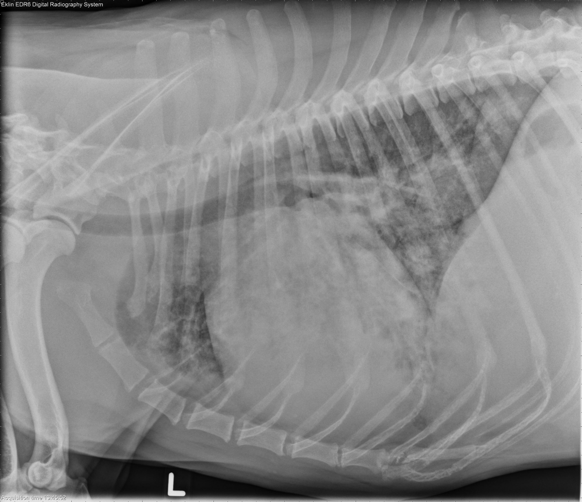
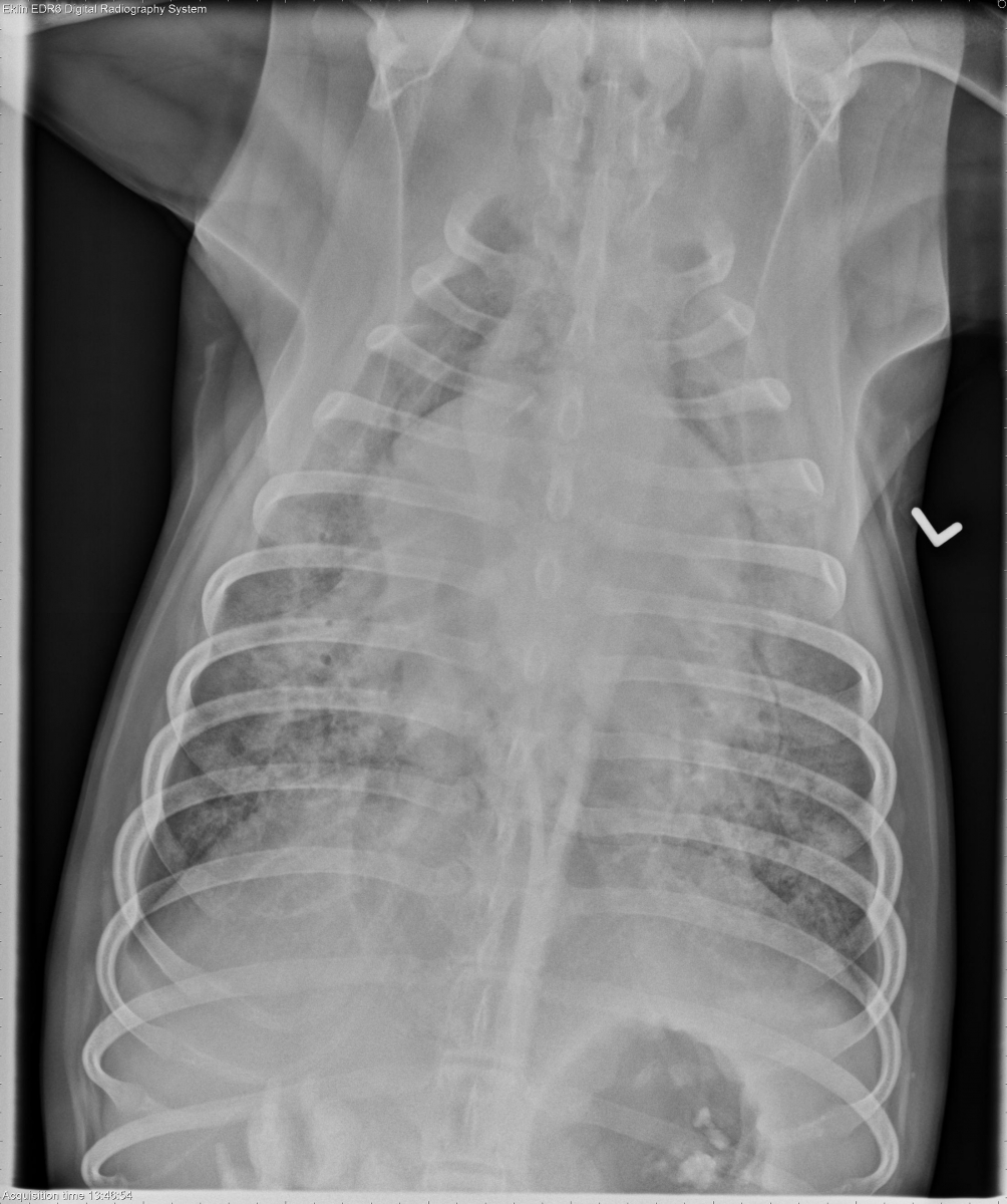
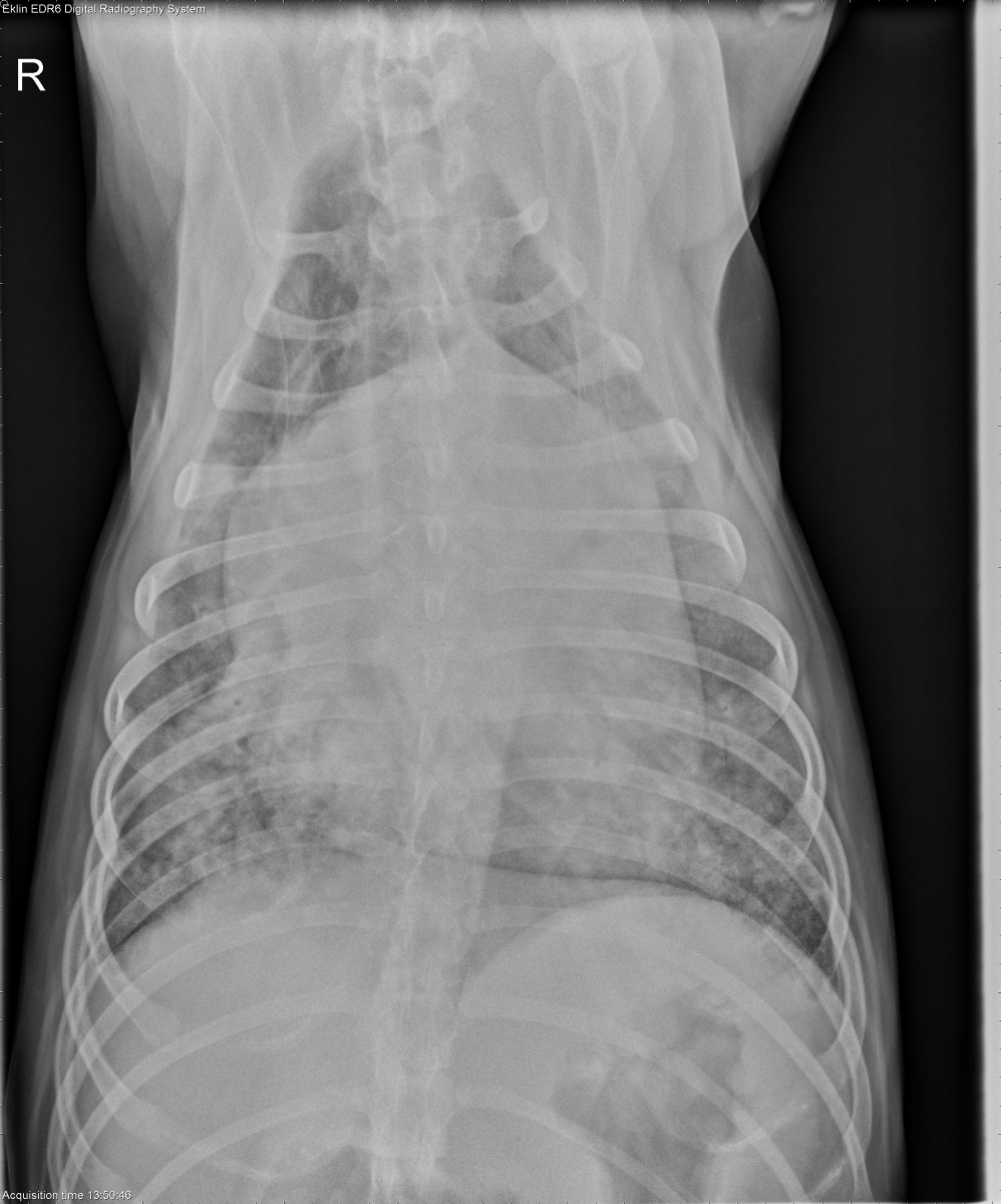
The cardiac silhouette is markedly enlarged with elevation of the trachea. The heart occupies more than half the thoracic width on the dorsoventral projection. The pulmonary vasculature appears enlarged in the right caudal lung lobe and normal in the remaining lung lobes. There is a diffuse, patchy interstitial to alveolar pulmonary pattern that is most pronounced in the right caudal lung lobe. The musculoskeletal structures appear normal. The peritoneal detail appears decreased.
Differential Diagnosis
Cardiomegaly with pulmonary edema and peritoneal effusion indicating left and right heart failure (valvular degeneration, dilated cardiomyopathy).
Diagnosis
Cardiomyopathy was diagnosed on echocardiography.
Discussion
The relatively normal pulmonary vasculature and dependent distribution of the pulmonary edema with a nodular appearance are typical of chronic heart failure and diuretic therapy.
cardiomegaly and heart occupies more than half of the thoracic cavity. Pulmonary edema is seen with mild plural effusion.