13 year old male neutered Husky with one month history of voice change, coughing, and trouble swallowing. What are your findings?


Findings
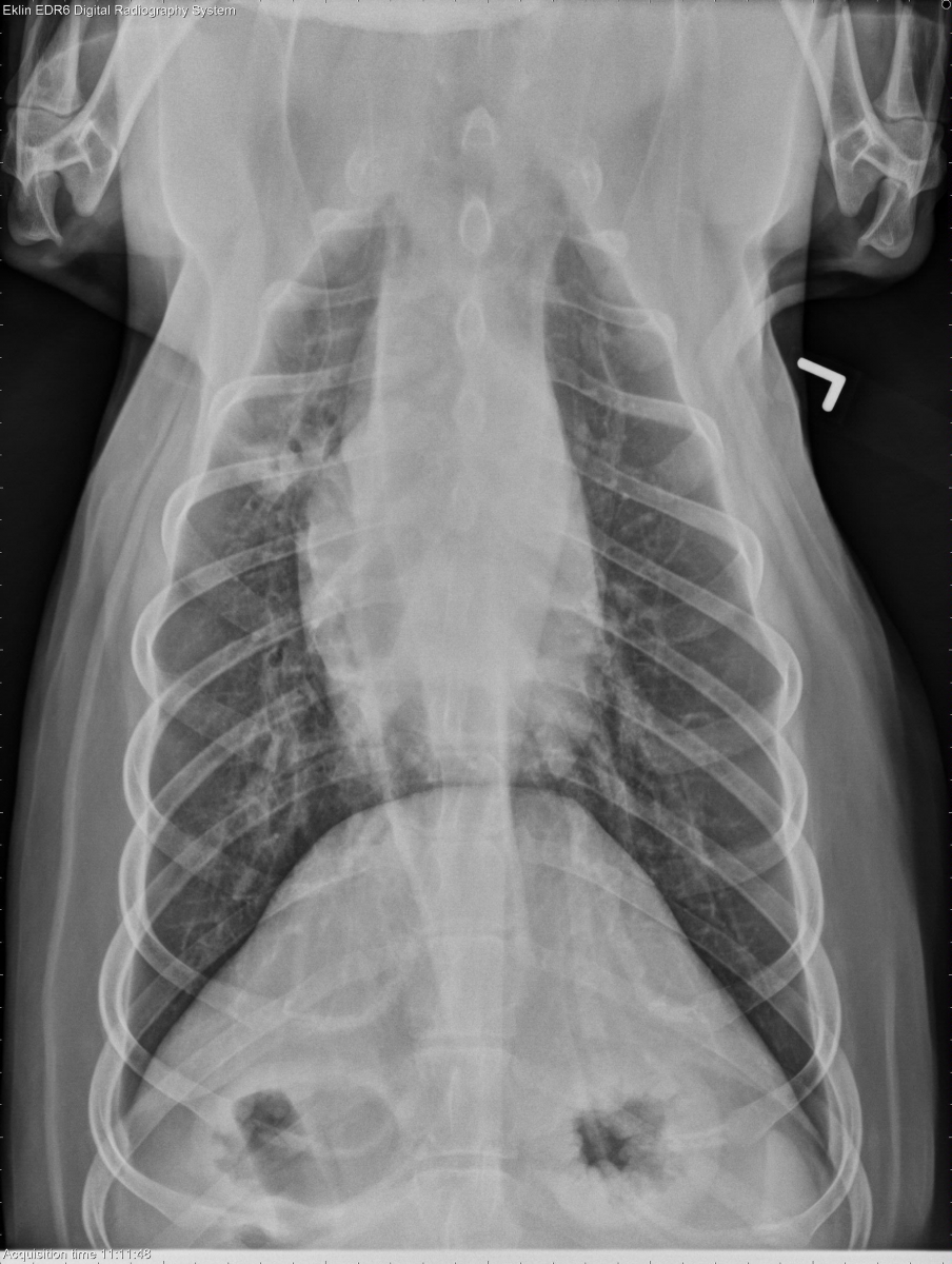
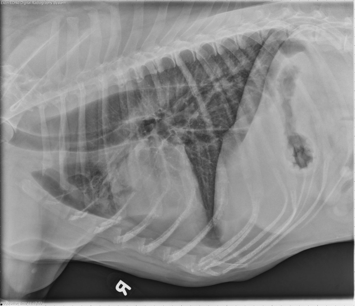
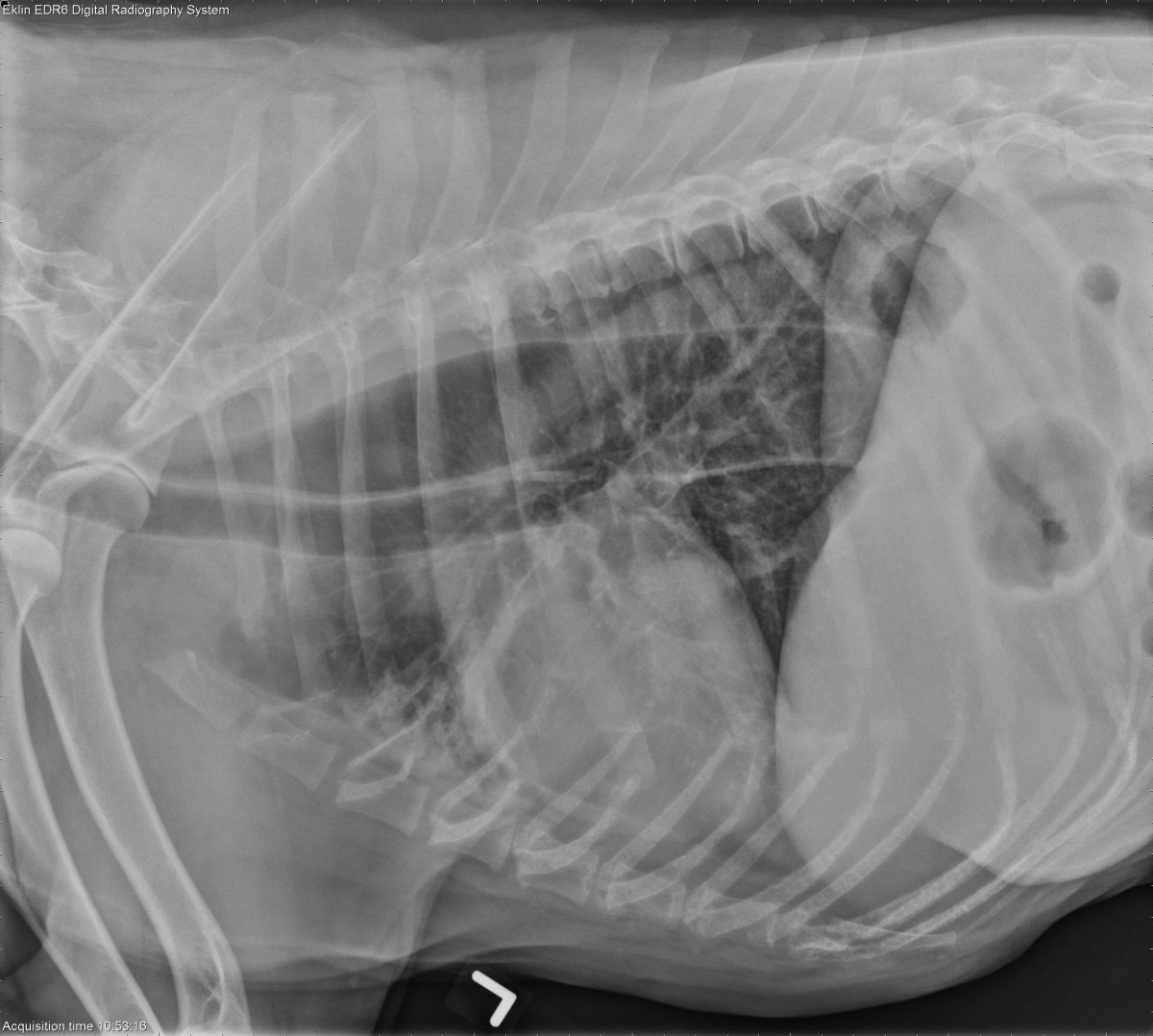
There is air in the laryngeal saccules on one image of the neck. The esophagus is diffusely dilated with air. There is an alveolar pattern in the right cranial lung lobe with an air bronchogram visible. The cardiovascular structures are normal.
Differential Diagnosis
- Megaesophagus
- Air in the laryngeal saccules can be associated with laryngeal paralysis
Diagnosis
- Neurogenic laryngeal and esophageal muscle atrophy causing laryngeal paralysis and megaesophagus with aspiration pneumonia
- No histologic abnormalities of the nerve were found
- Most likely idiopathic in origin
References
- Kitshoff, AM., BV Goethem, L Stegen, P Vandekerckhove, H de Rooster. 2013. Laryngeal paralysis in dogs: An update on recent knowledge. Journal of the South African Veterinary Association 84(1):1-9.
On thorax lateral projection, gas dilated esophagus displacing trachea ventrally. On cranial to cardiac silhouette, there seems the most remarkable bronchial pattern with small pulmonary artery and vein. On VD projection, more bronchial pattern on the right hemi-thorax than the counterpart. Gas-filled esophagus demarcation is observed mediastinum, as well.
DX – Megaesophagus causing secondary aspiration pneumonia.
Initial TX – Broadspectrum AB for a month with nursing therapy including upright position feeding with fluidic or proper consistency of food. Coupage, hydration and nebulization with oxygenation. If no response to AB therapy within a week, culture and change AB.
I guessed that there is a problem with larynx due to voice tone change and swallowing disability but failed to point it out.
1. If there are gas opacity within the larynx structure, is it a pathognomonic sign for the everted laryngeal saccule?
2. In some cases I don’t see any structures within larynx but soft cartilages while ,in some cases, I see soft tissue structure only. Both situations are normal?
Always thank you in advance.
On one of the lateral views of neck, there is an ovoid soft tissue opaque mass in the nasopharynx which is however not seen on the other lateral view of neck. Is it normal?
Thank you in advance.