This week’s case is a 4-year-old female neutered Labrador Retriever with fever of unknown origin. What are your findings?
Findings
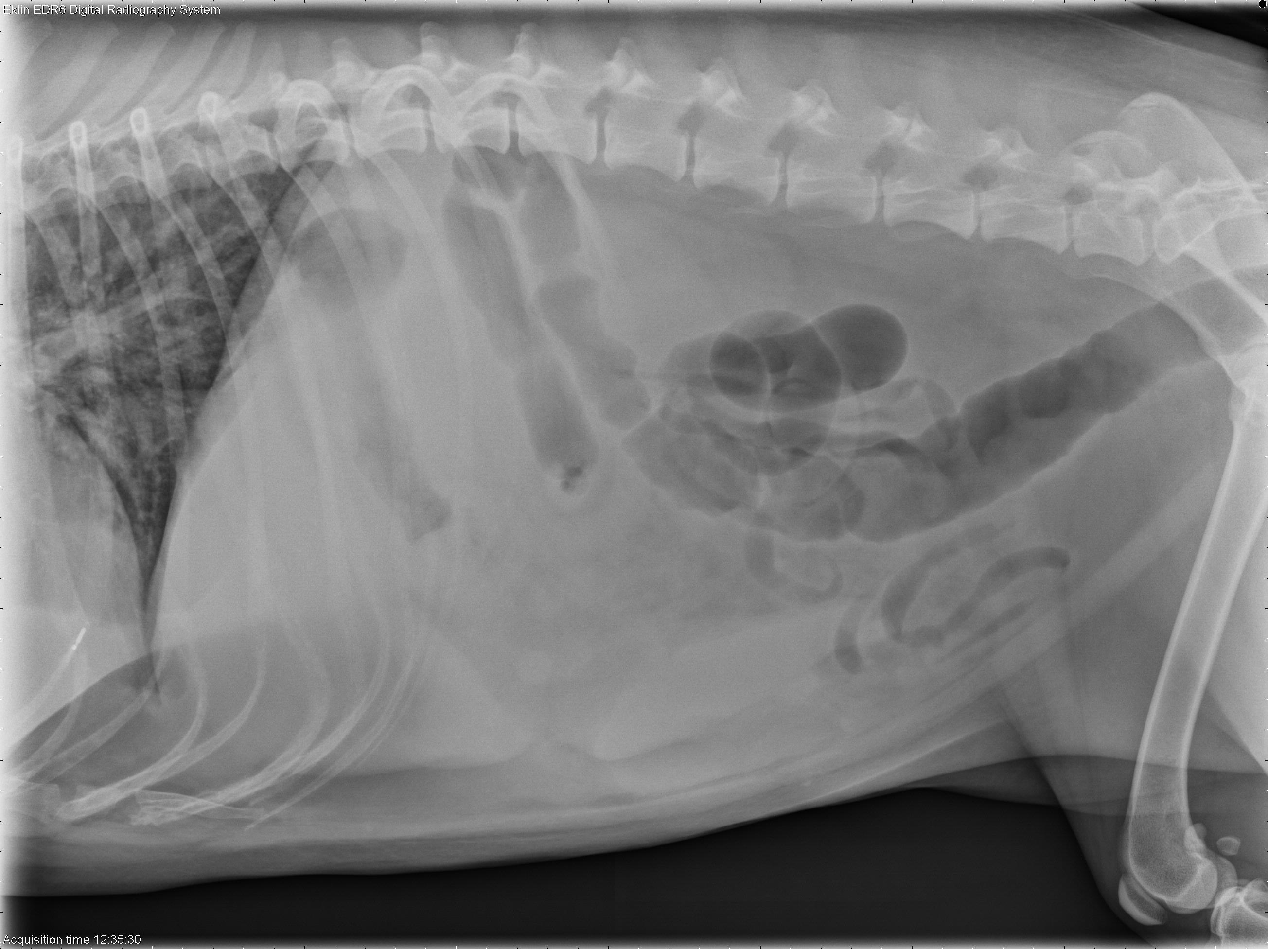
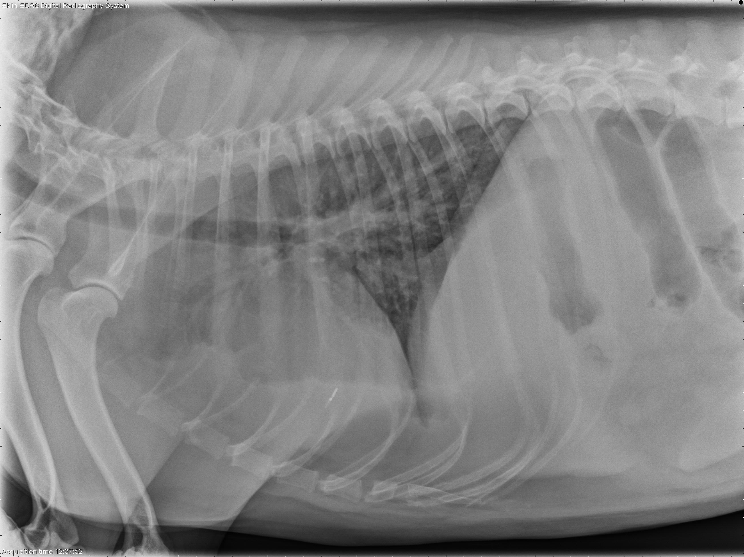
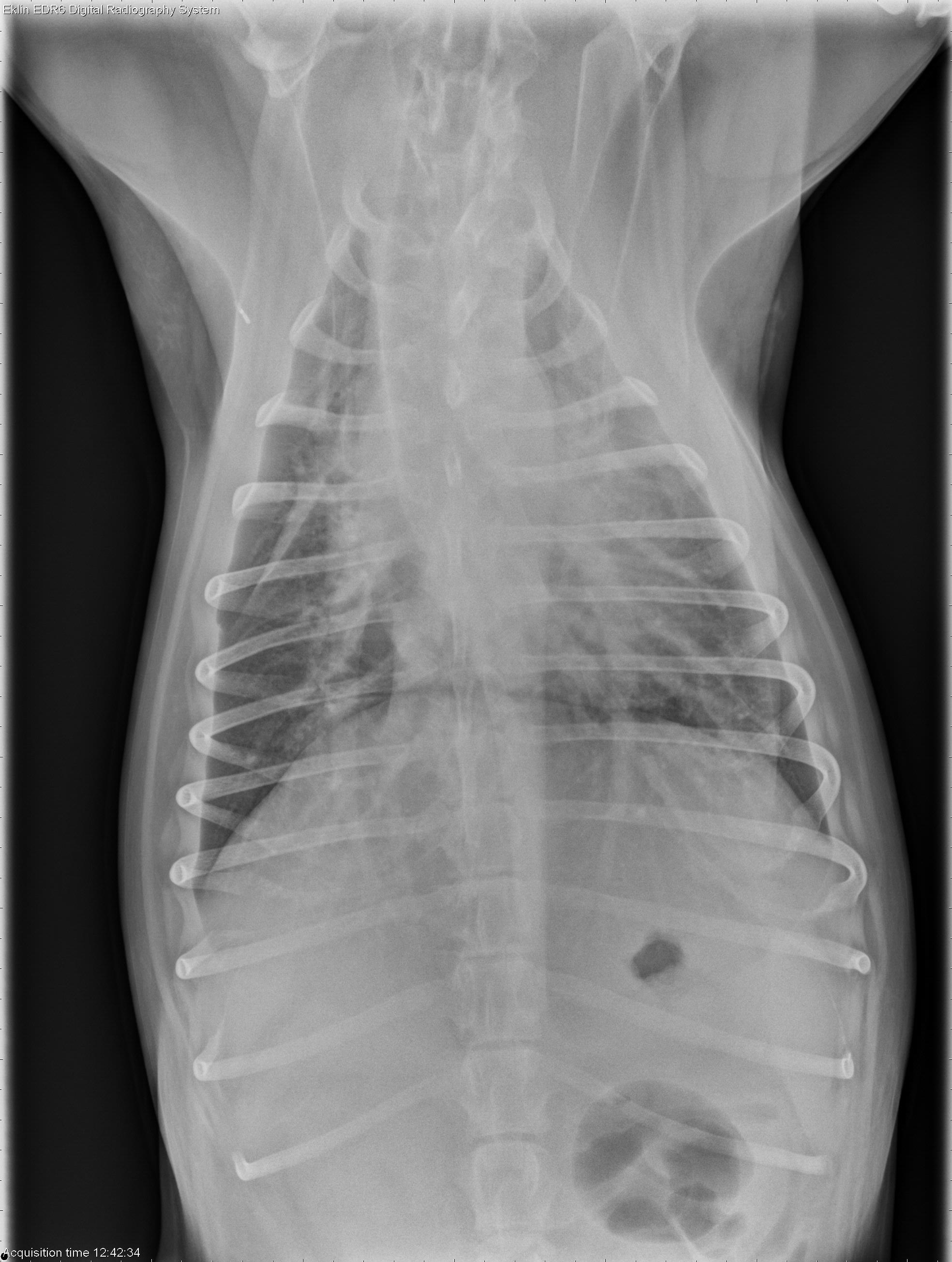
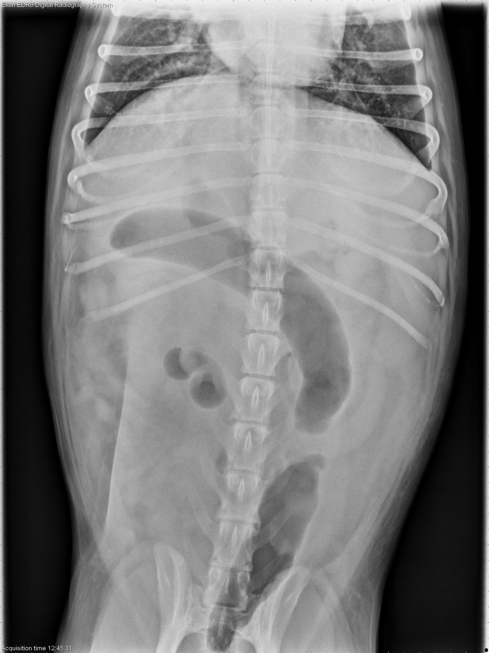
Thorax: There is increased soft tissue opacity in the pleural space with retraction of the lung lobes and partial silhouetting with the cardiac silhouette. There is a patchy bronchointerstitial pattern in the lungs. The cardiovascular structures appear normal.Abdomen: The abdomen is distended and there is poor peritoneal detail. The liver and spleen are enlarged with rounded borders. The colon is moderately dilated with gas. The small intestine appears normal. The urinary tract is not well visualized.
why is it pulmonary edema and not pleural effusion? In the lateral projection of the chest it appeared to be pleural effusion
It is both. That’s why the bicavitary effusion and soft tissue opacity in the pleural space with retraction of the lung lobes and partial silhouetting with the cardiac silhouette was mentioned.
thanks
Another doubt would be in the Dorso-ventral projection of the chest the vessels were very evident in the left caudal lobe. Is this just an effect of the positioning or does it indicate a change?