The first case of 2019 is a 7-year-old FS Domestic Medium-hair cat presented with dypsnea. What are your findings?
Findings
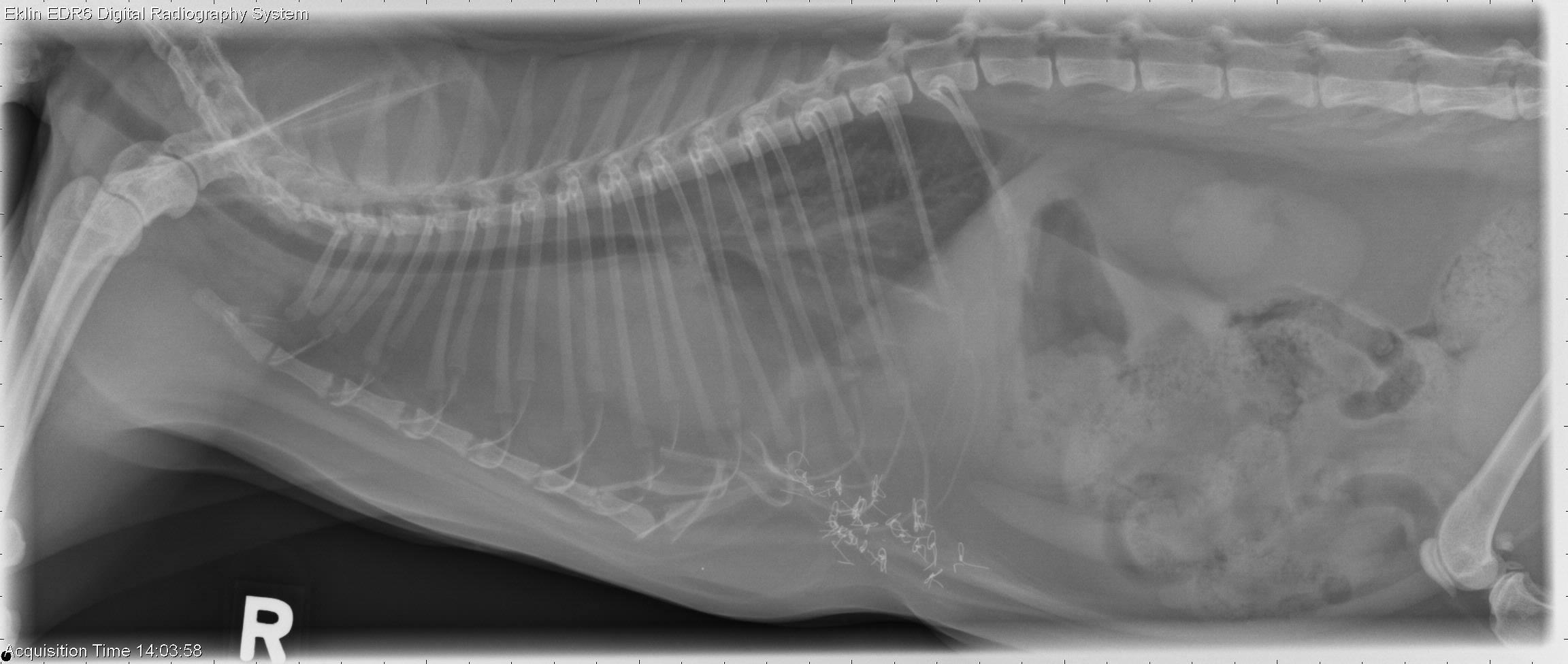
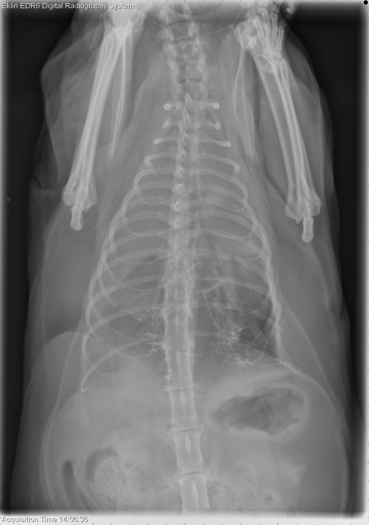
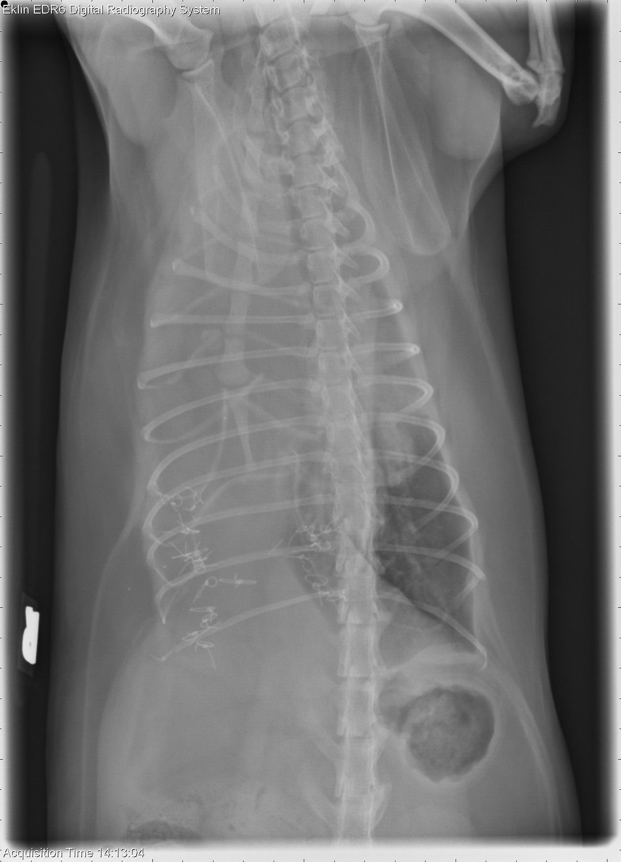
There is a significant increase in fat opacity within the cranioventral thorax. The heart is caudodorsally displaced. The xyphoid is displaced dorsally and multiple metallic sutures are identified within the ventral abdomen. The cardiac silhouette is difficult to visualize on the VD projection due to overlying fat. The right diaphragmatic crus is difficult to visualize on the ventrodorsal projections.
Differential Diagnosis
- Diaphragmatic rupture
- Sutures are likely secondary to previous diaphragmatic repair
Diagnosis
- A diaphragmatic rupture was confirmed at surgery and was repaired.
Hey,
I don’t understand why a significant increase in fat opacity within the cranioventral thorax has diaphragmatic hernia as main DD
Could you please explain ?
Yours,