This week’s case is a 3-year-old male neutered Labrador Retriever with one-month history of progressive left thoracic limb lameness. What are your differential diagnoses?
Findings
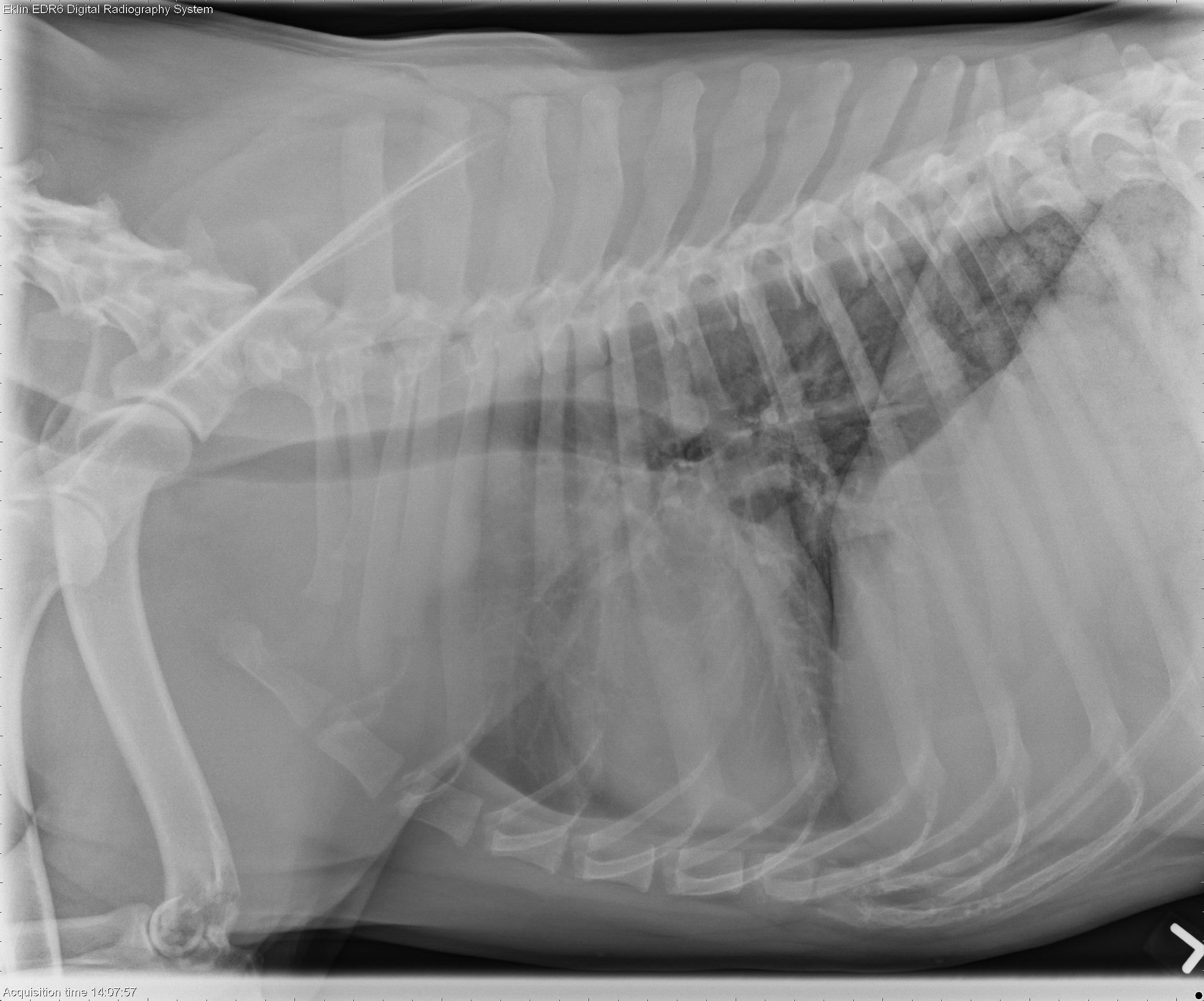
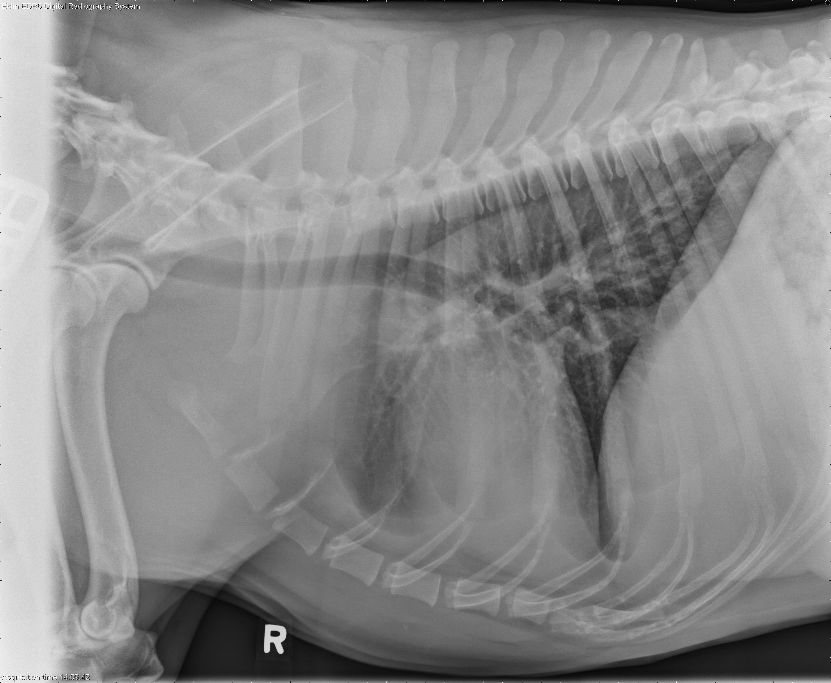
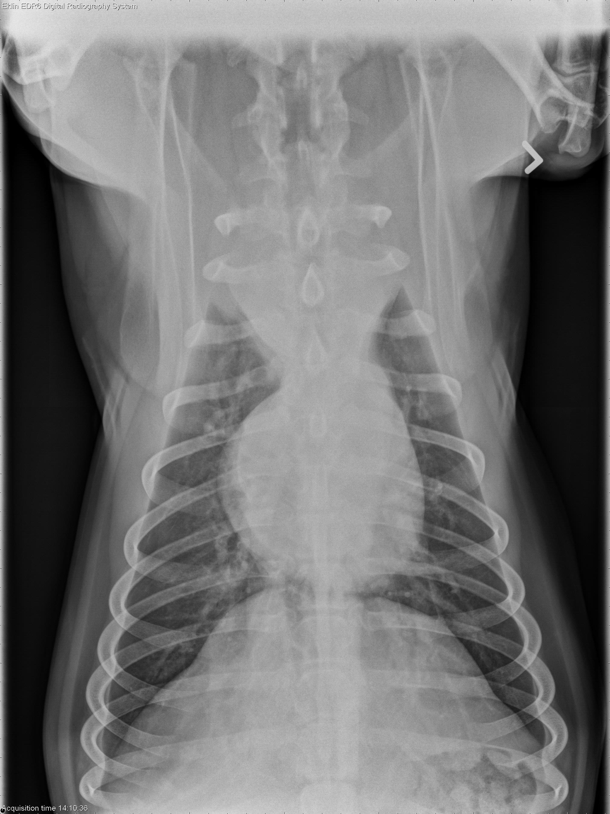
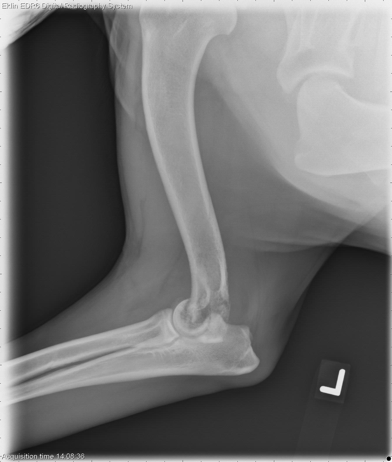
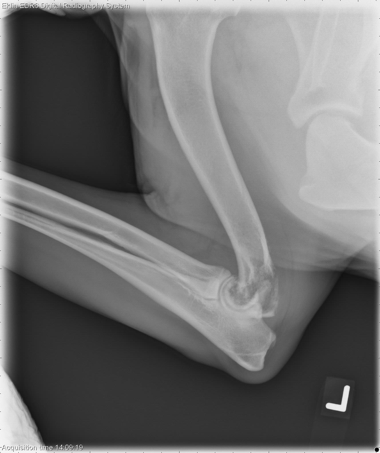
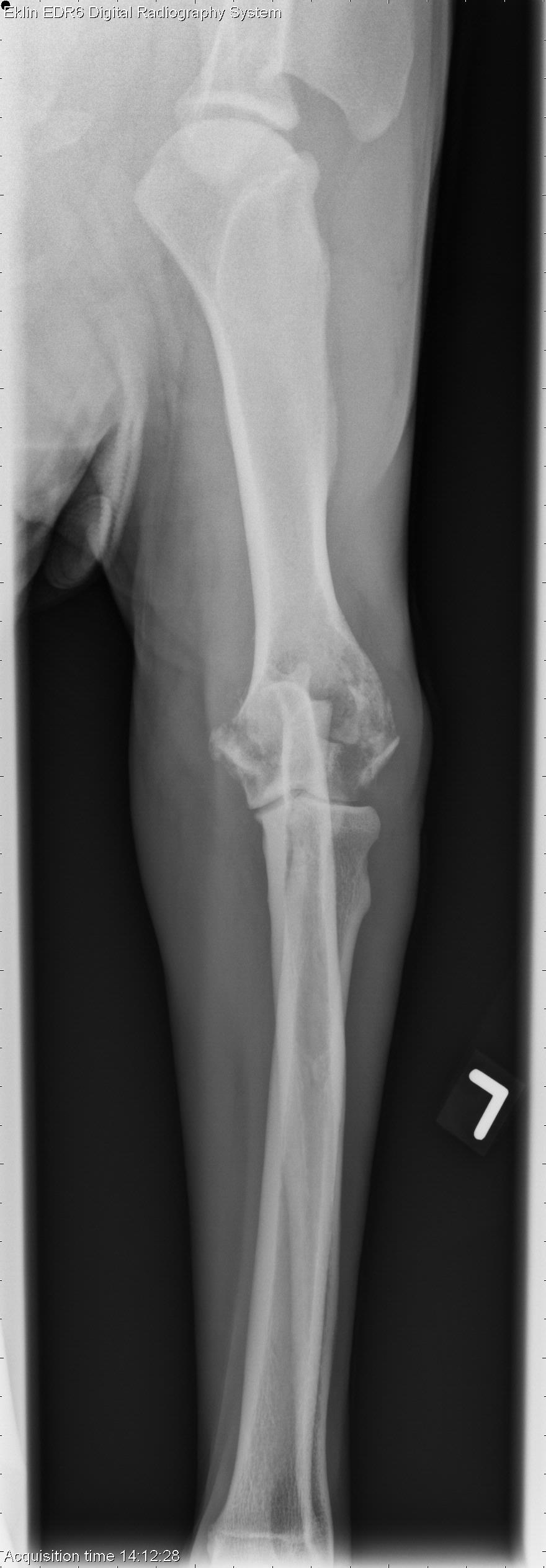
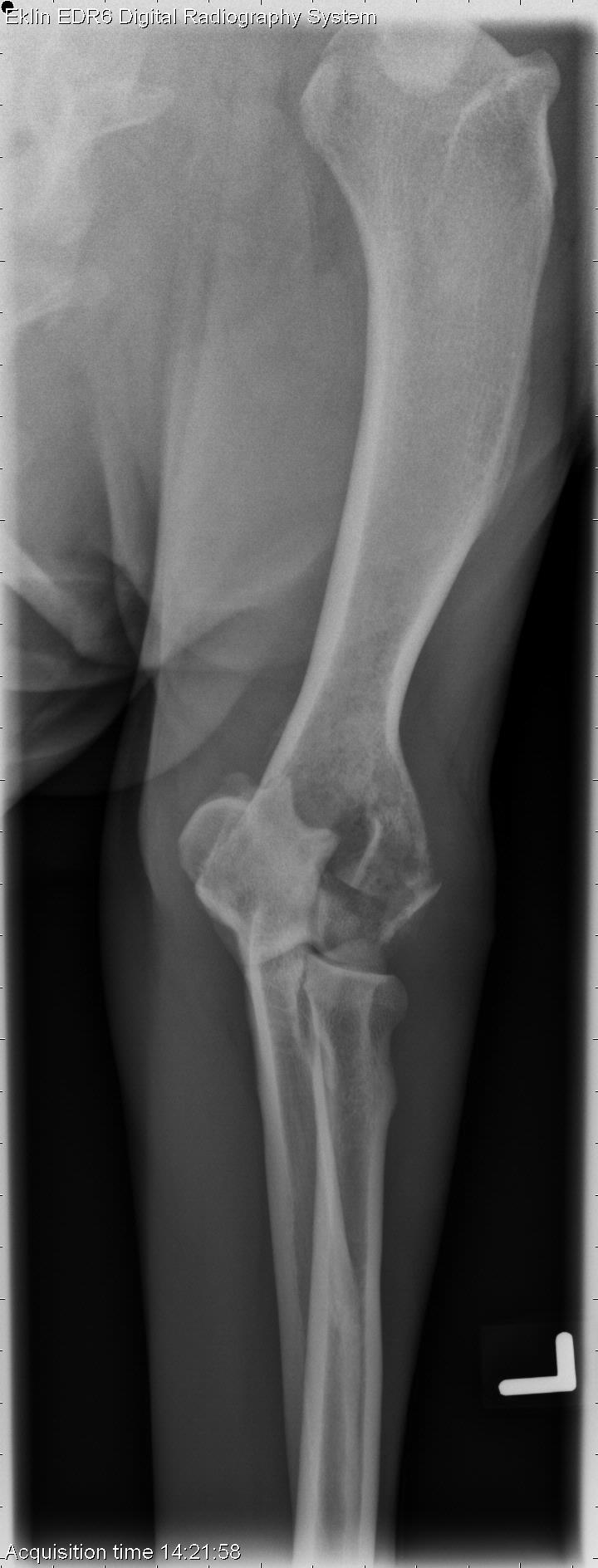
Thorax: There is increased soft tissue opacity in the cranial thorax on the lateral projections, and widening of the mediastinum on the dorsoventral projections. The trachea is elevated on the lateral projections. The cardiovascular structures are normal. There is a mild diffuse interstitial pattern throughout the lungs.Left thoracic limb: Neutral, flexed lateral, craniocaudal, and craniocaudal oblique projections of the left elbow are available for review. There is osteolysis of the humeral condyle and disruption of the cortex on the lateral projections. On the craniocaudal projections, a fracture line is visible traversing the lateral epicondyle with discontinuity of the cortex.
Differential Diagnosis
- Cranial mediastinal mass (lymphoma, thymoma)
- Aggressive bone lesion of the left humerus with pathologic fracture (metastatic neoplasia, osteomyelitis less likely)
Diagnosis
- Disseminated T-cell lymphoma with metastasis to left humerus
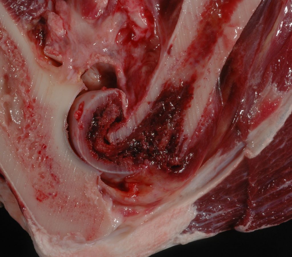
- Left thoracic limb was amputated, pathologic fracture confirmed
If the data is available, what color was this Labrador?
Thank you!
Dr. Ben Daggett
Tug Hill, NY