This week’s case is a 10-year-old female neutered Greyhound with 2-week history of decreasing appetite and energy. It’s a challenging one. What are your findings?
Findings
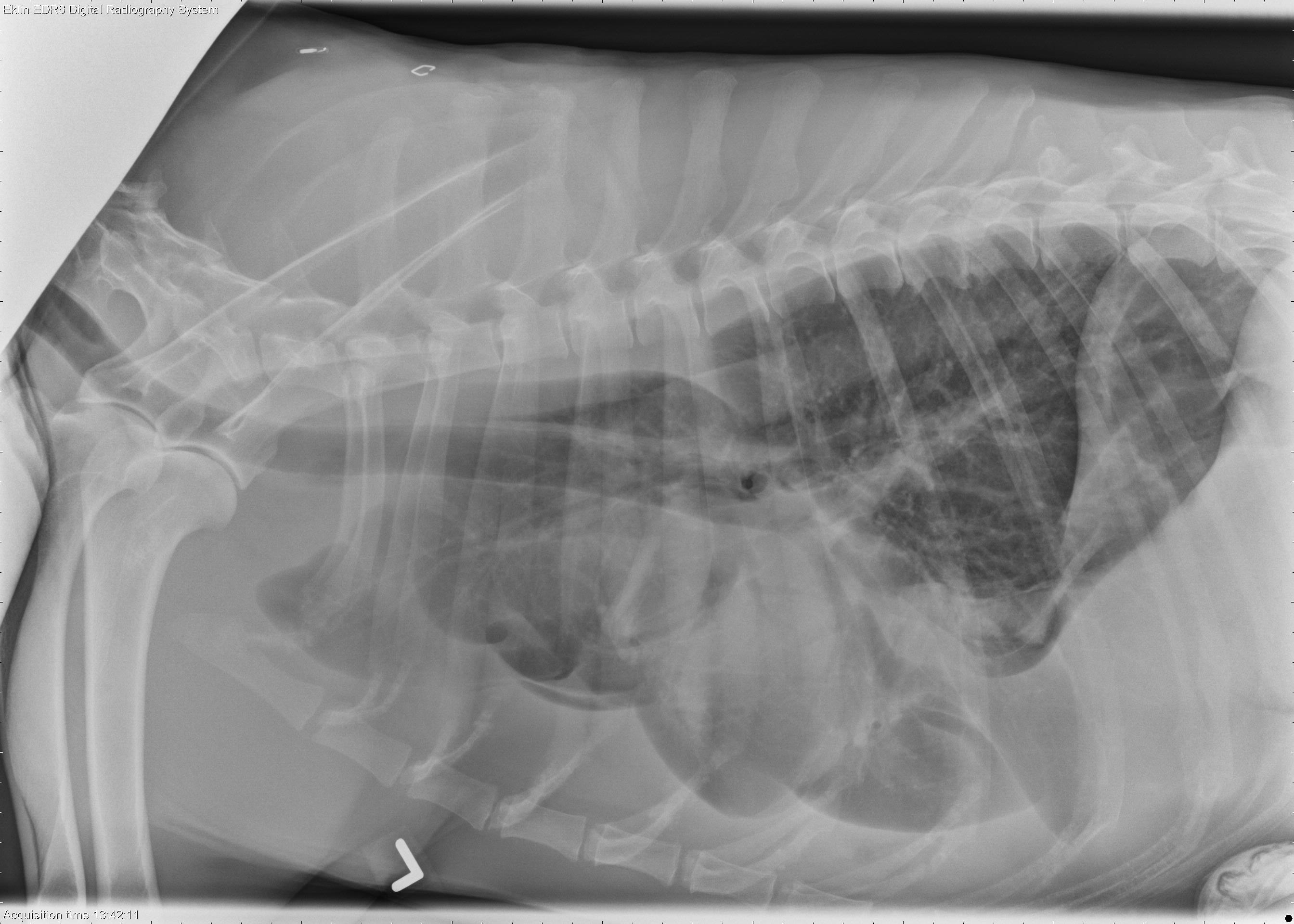
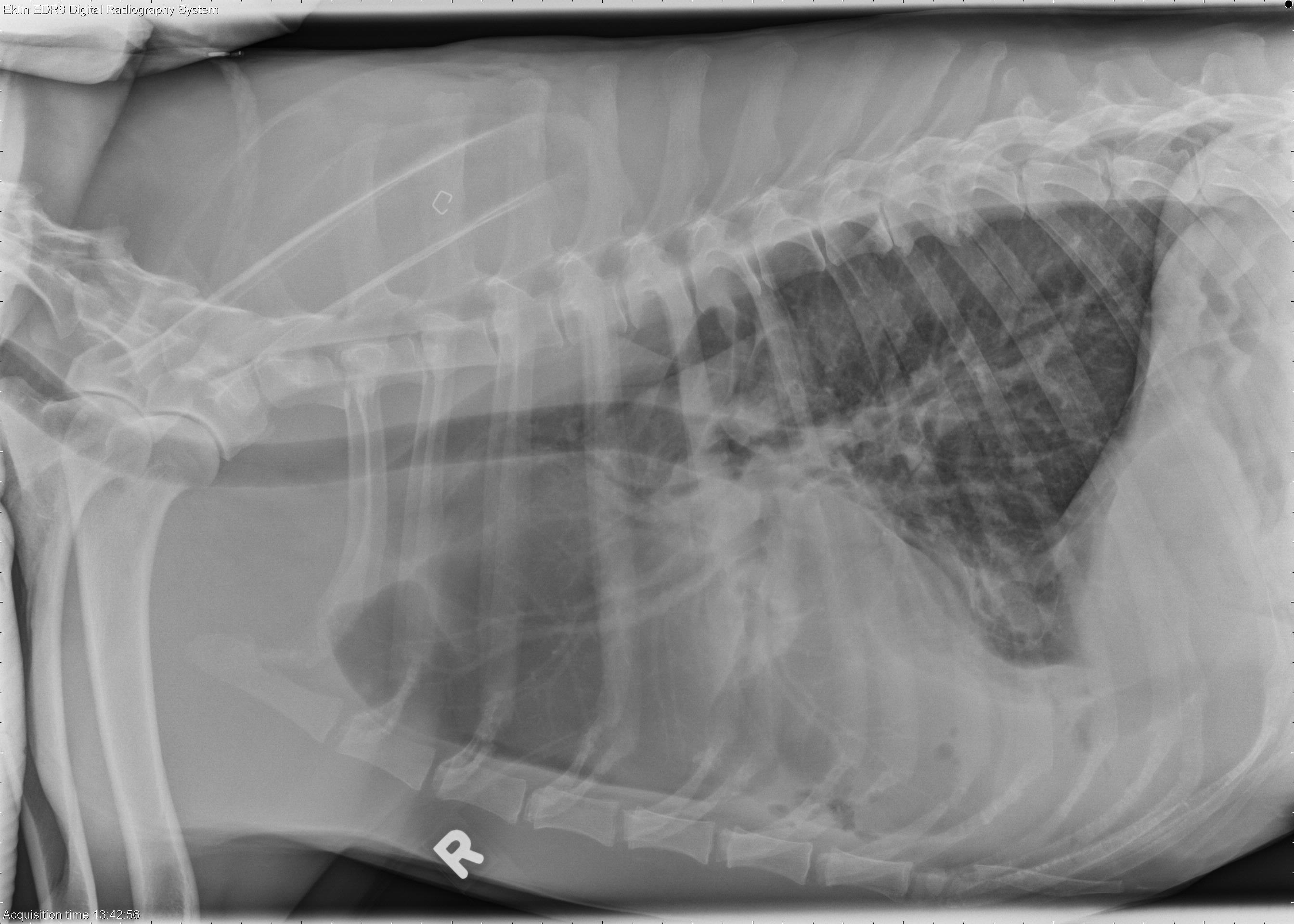
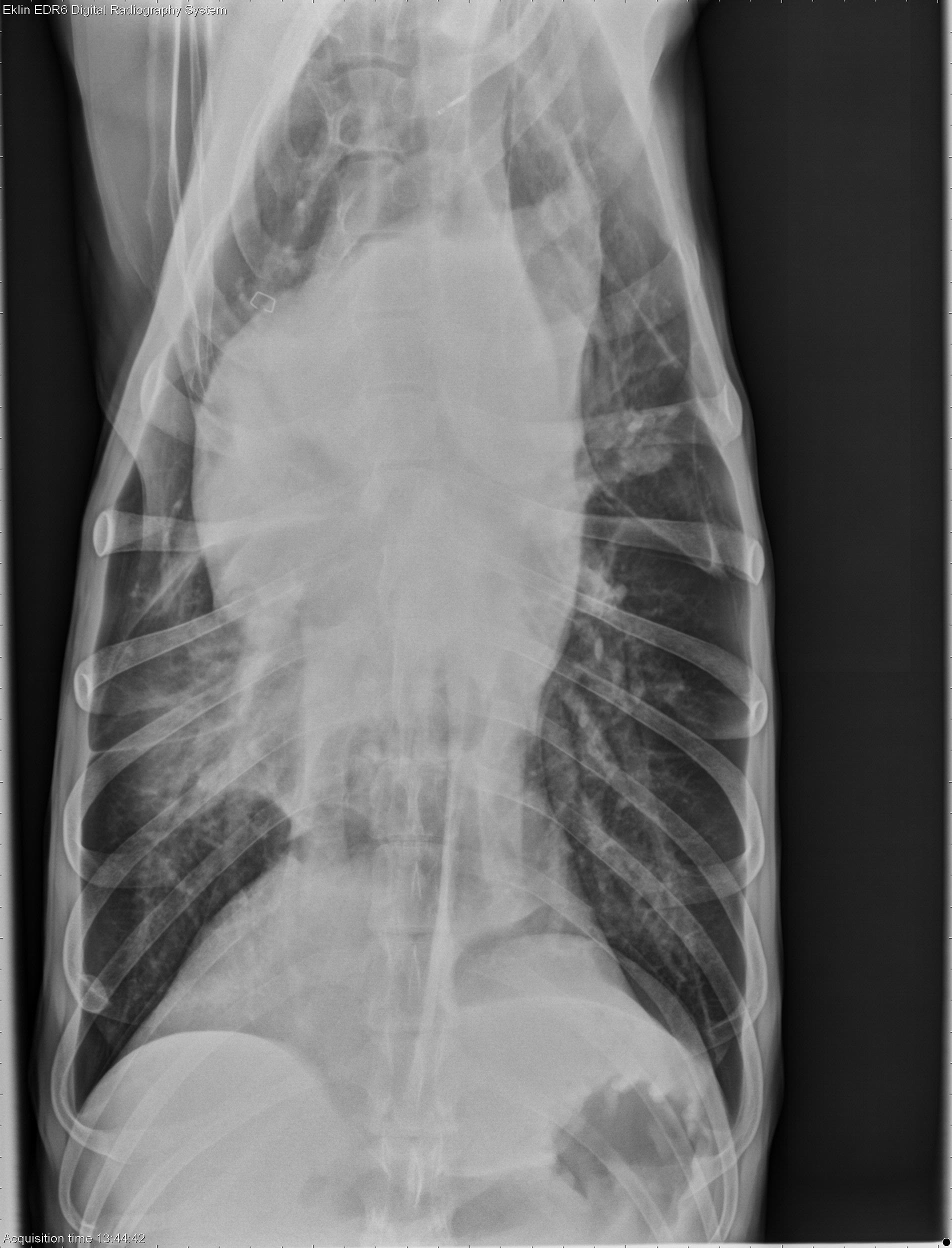
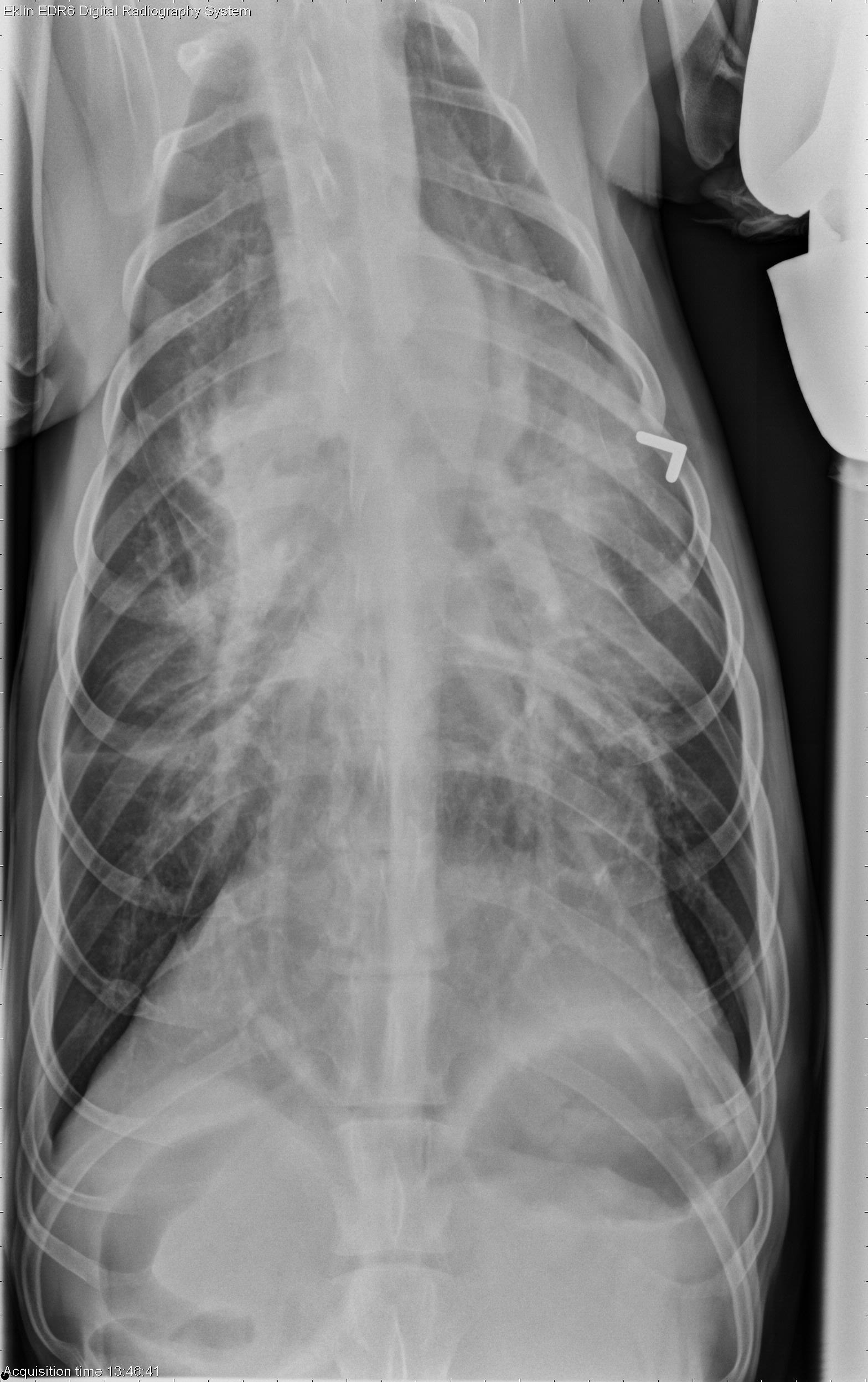
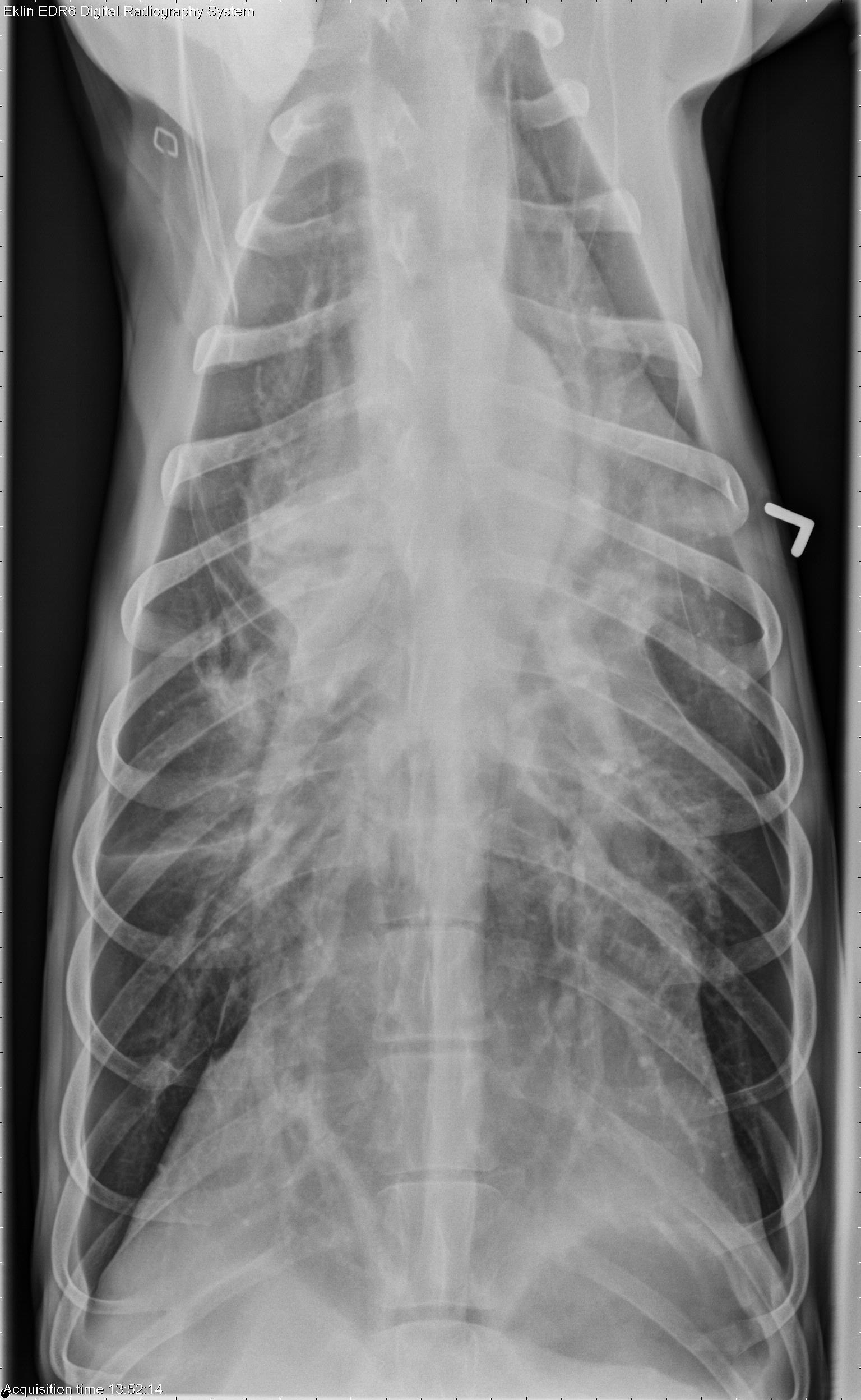
Left and right lateral, ventrodorsal, and dorsoventral radiographs are available for review. There is moderate pleural effusion present with rounding and retraction of the lung lobes. In addition, there is a mass effect between the cardiac silhouette and the diaphragm in the caudoventral thorax. This appears to be a mass in the right caudal lung lobe on the ventrodorsal projection. The region of the accessory lung lobe also appears more opaque. There are small gas bubbles visible on the right lateral projection which may be in the pleural space or within the mass. There is a triangular shaped projection at the left cranial cardiac silhouette on the ventrodorsal projection which may represent a reverse fissure line, indicating fluid in the mediastinum. The crannial a mediastinum appears widened on the dorsoventral projection.Differential Diagnosis
- Suspected right caudal lung lobe mass, +/- accessory lobe with pleural effusion (Pulmonary abscess, pneumonia due to migrating grass awn, with pyothorax). Other differentials could include granuloma, neoplasia with secondary effusion but are less likely.
- Mediastinal widening – mediastinal fluid, abscessation, lymphadenopathy, mass
Diagnosis
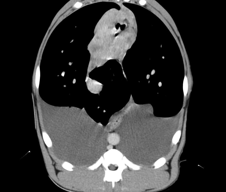
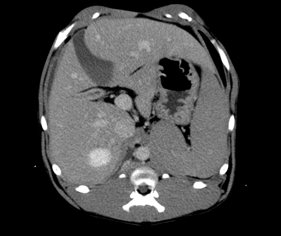
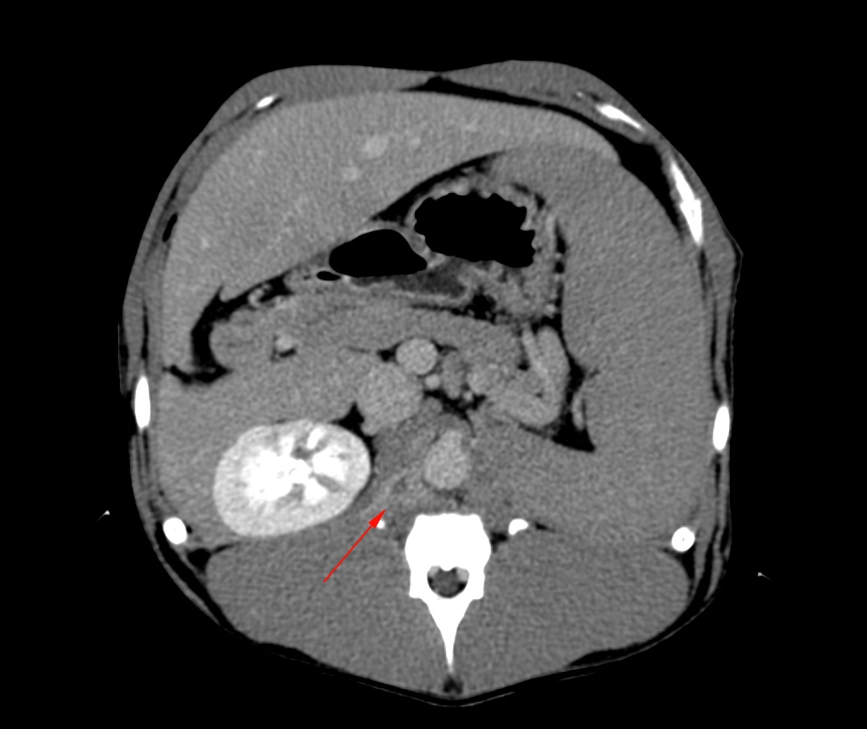
- On CT examination, the suspected lung lobe mass was found to be an abscess in the caudal mediastinum. There were multiple areas of focal consolidation at the periphery of the lung, suspicious of foreign body migration. The contrast enhancing tracts and gas extended into the retroperitoneal space (CT images)
- The mediastinal tissue was resected at surgery, along with an adherent portion of the left caudal lung lobe. A laparotomy was also performed to excise the abscessed tissue in the epaxial muscles.
- Pyothorax, pyogranulomatous mediastinitis, diffuse consolidating pneumonia with bacteria and fibrosis. Presumed secondary to inhaled grass awn migration.
why did you suspect a mass and not an hernia?