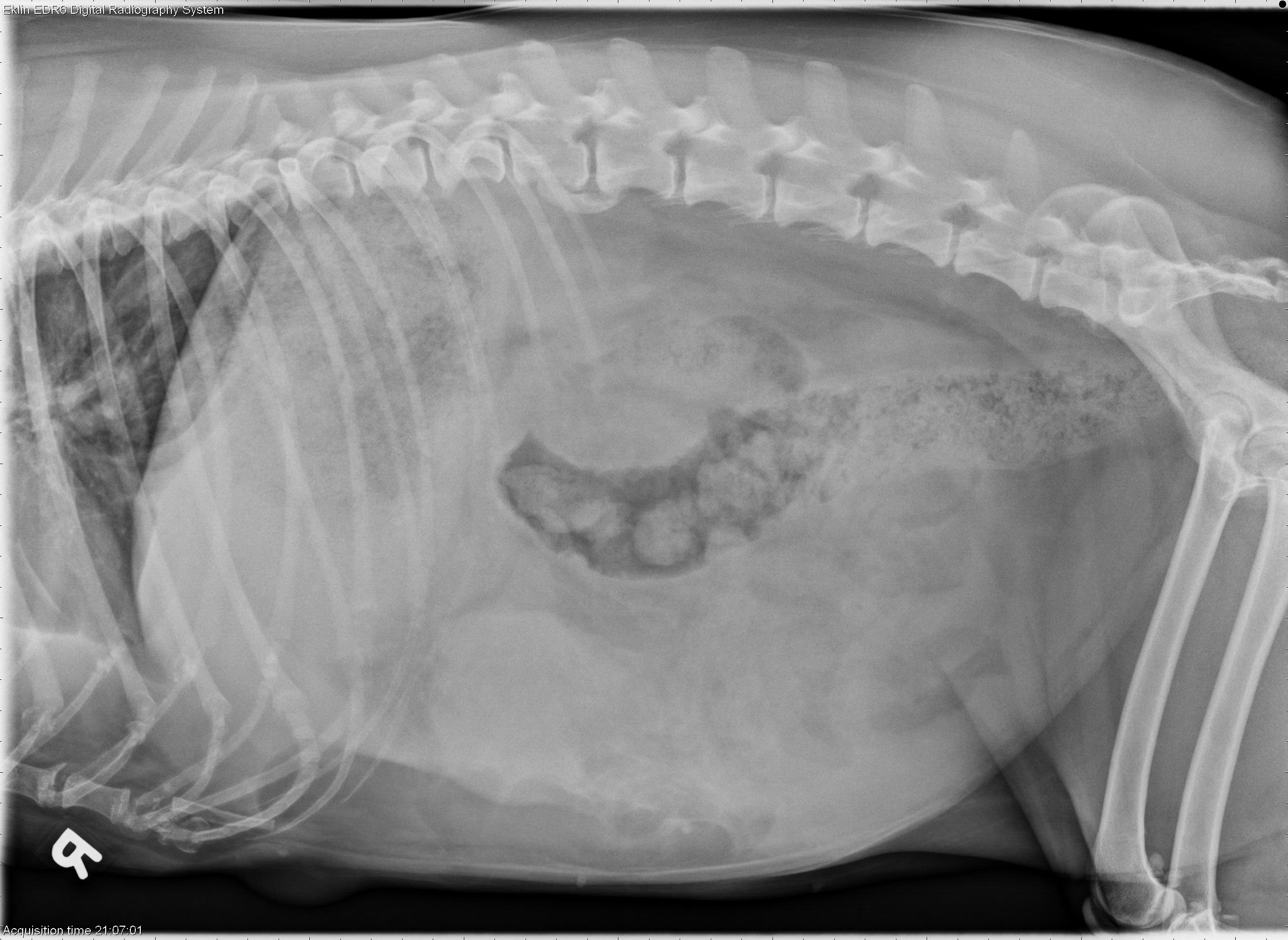
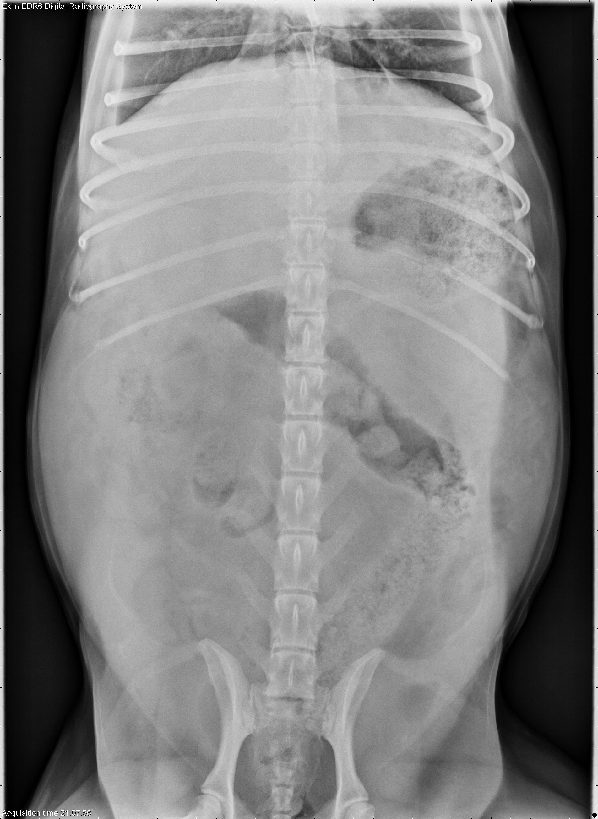
Happy new year! Today’s case is a 12-year-old female spayed Labrador-Border Collie cross presented to Emergency Services after becoming recumbent and having difficulty breathing this evening. What are your findings?
Teaching and learning about veterinary diagnostic imaging.
Happy new year! Today’s case is a 12-year-old female spayed Labrador-Border Collie cross presented to Emergency Services after becoming recumbent and having difficulty breathing this evening. What are your findings?
Is there also a pleural effusion seen at the edge of the lateral view? Or is that just fat?