Today’s case is a 9 year old male neutered Golden Retriever with a dry, retching cough of 6 months duration. Initially treated with antibiotics, cough improved but was still intermittently present. In the last three weeks, the dog has progressed to coughing 2-3 times per hour, which has improved to only 2-3 times a day with reinstitution of antibiotic therapy.
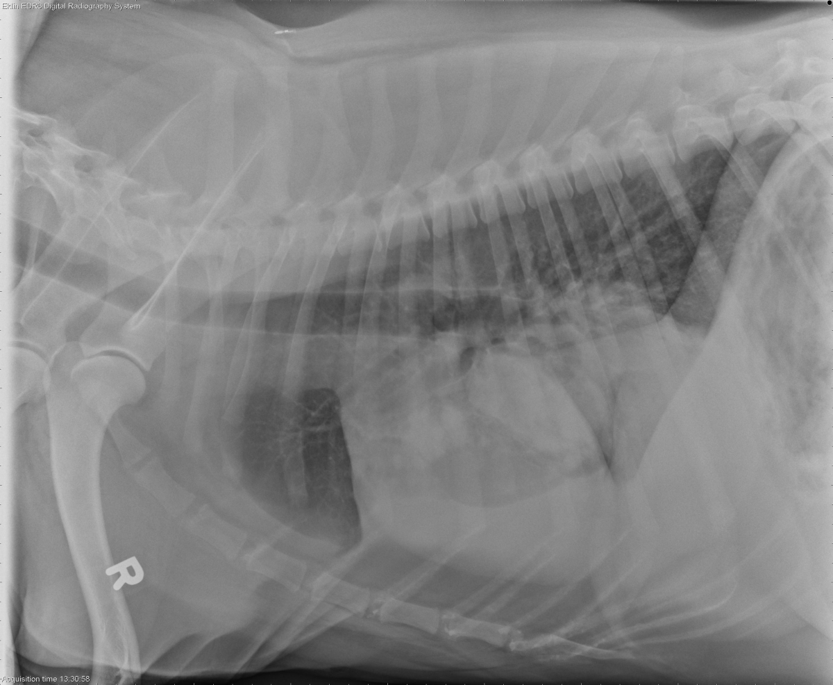
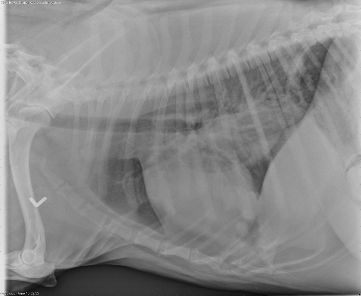
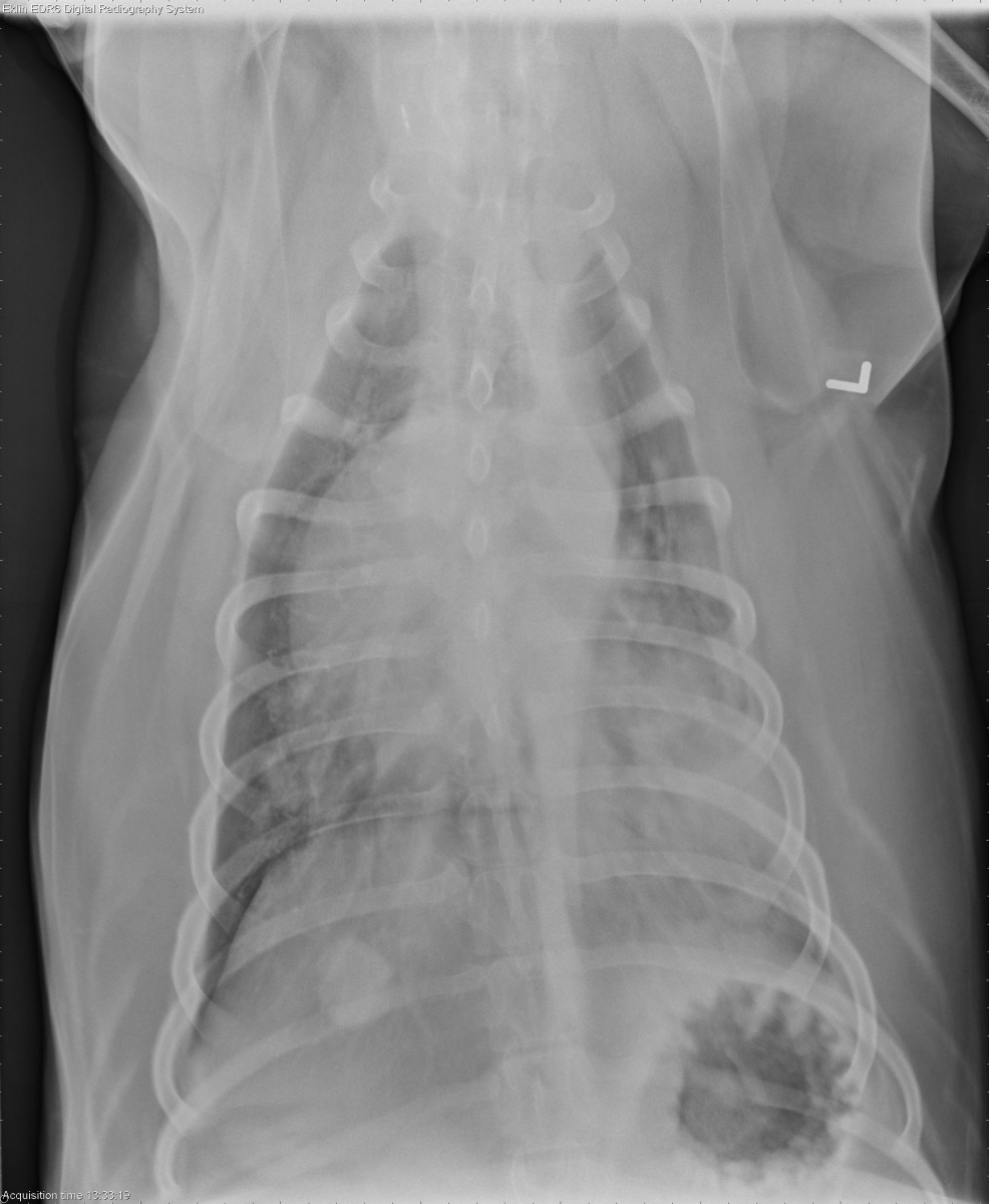
Findings
Three well defined, 2-3 cm soft tissue masses are observed on the lateral and DV projections. Multiple smaller, poorly defined soft tissue nodules are also observed on the right lateral projection. There is a severe alveolar pattern in the dependent portion of the left caudal lung lobe. This may represent a poorly defined mass. A small amount of gas is present in the esophagus dorsal to the carina. The cardiovascular structures appear within normal limits.
Differential Diagnosis
- primary lung tumor (carcinoma) in the left caudal lobe with metastasis
- multiple metastases from distant tumor
- less likely-infectious/inflammatory disease
Diagnosis
Pulmonary carcinoma (fine needle aspirate)
Discussion
The abdominal ultrasound was normal. The larger mass in the left caudal lung lobe is most likely the primary tumor with metastasis to the other lung lobes. Carcinomas can cause ill-defined masses as well as diffuse interstitial metastases. The mass was visible through the diaphragm on abdominal ultrasound, as well as through the intercostal spaces on thoracic ultrasound. Fine needle aspirates were obtained through this window.
Is it legit to say that fat tissue opacity in the pleural space is related to obesity?
Yes, there is fat in the mediastinum ventrally that causes some retraction of the lung lobes on the lateral projections.