This week’s case is a 2-year-old male-neutered Shih Tzu presented for 1-month history of coughing and weight loss. Take a look and post your comments!
Findings
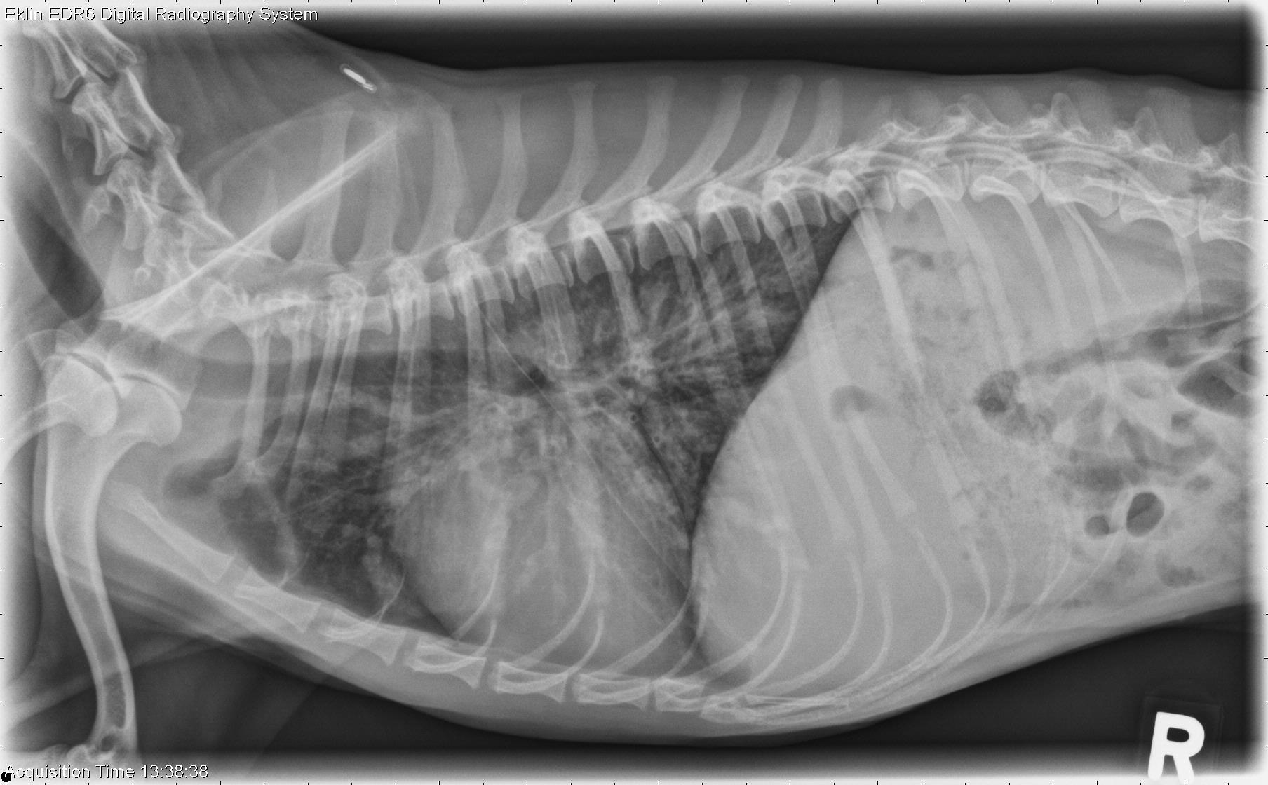
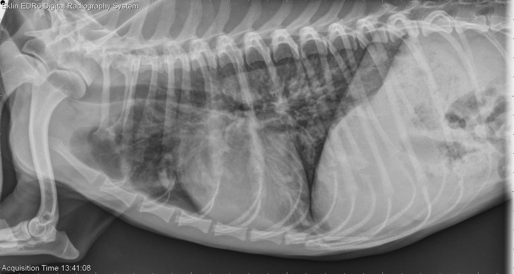
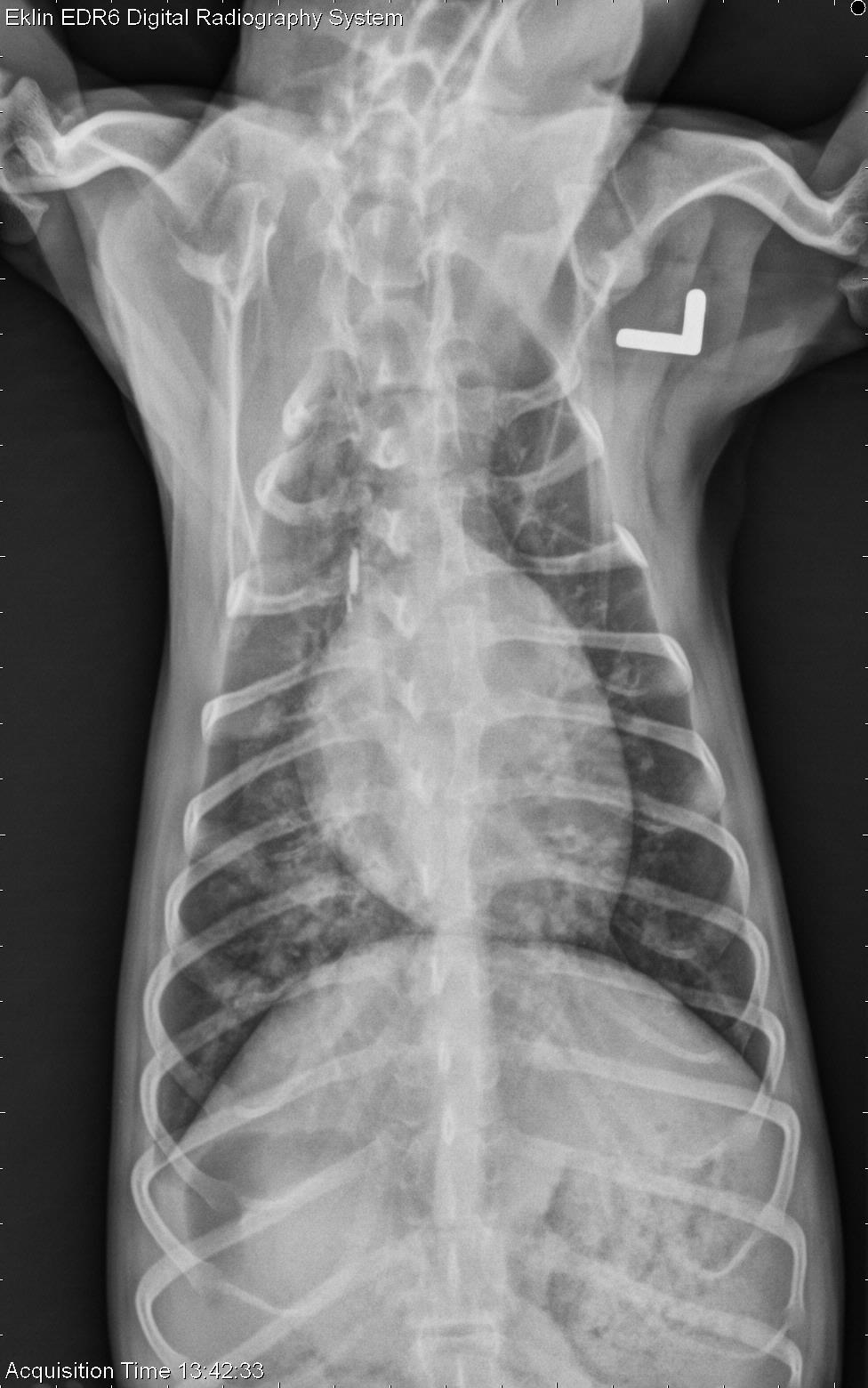
There is a severe bronchial pattern throughout the lungs. Soft tissue dense material lines multiple airways. The bronchi closer to the hilus have a “doughnut” appearance, and more distally fluid filled bronchi appear nodular. A thin pleural fissure line seen crossing the thorax on the right lateral view. The cardiovascular structures are within normal limits. Within the viewable abdomen there is decreased serosal detail which is likely related to the thin body condition of the patient.
Differential Diagnosis
Severe bronchial pattern consistent with bronchitis, with inflammation or debris lining multiple airways. This is most likely most likely immune mediated in orgin (eosinophilic bronchopneumopathy) however infectious inflammatory airway disease should be ruled out.
Diagnosis
- Eosinophilic bronchopneumopathy
- On bronchoscopic examination, there was mucus diffusely distributed in the airways. The bronchial lavage and cytology showed marked, mixed inflammation which was primarily eosinophilic.
[…] http://www.veterinaryradiology.net/3222/2-year-old-shih-tzu/ […]