Today’s case is a 3 month old female Husky with regurgitation since being weaned from her mother. What are your findings?
Findings
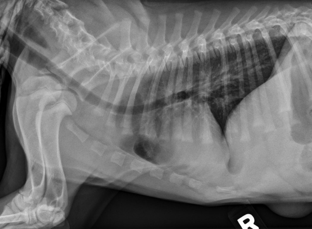
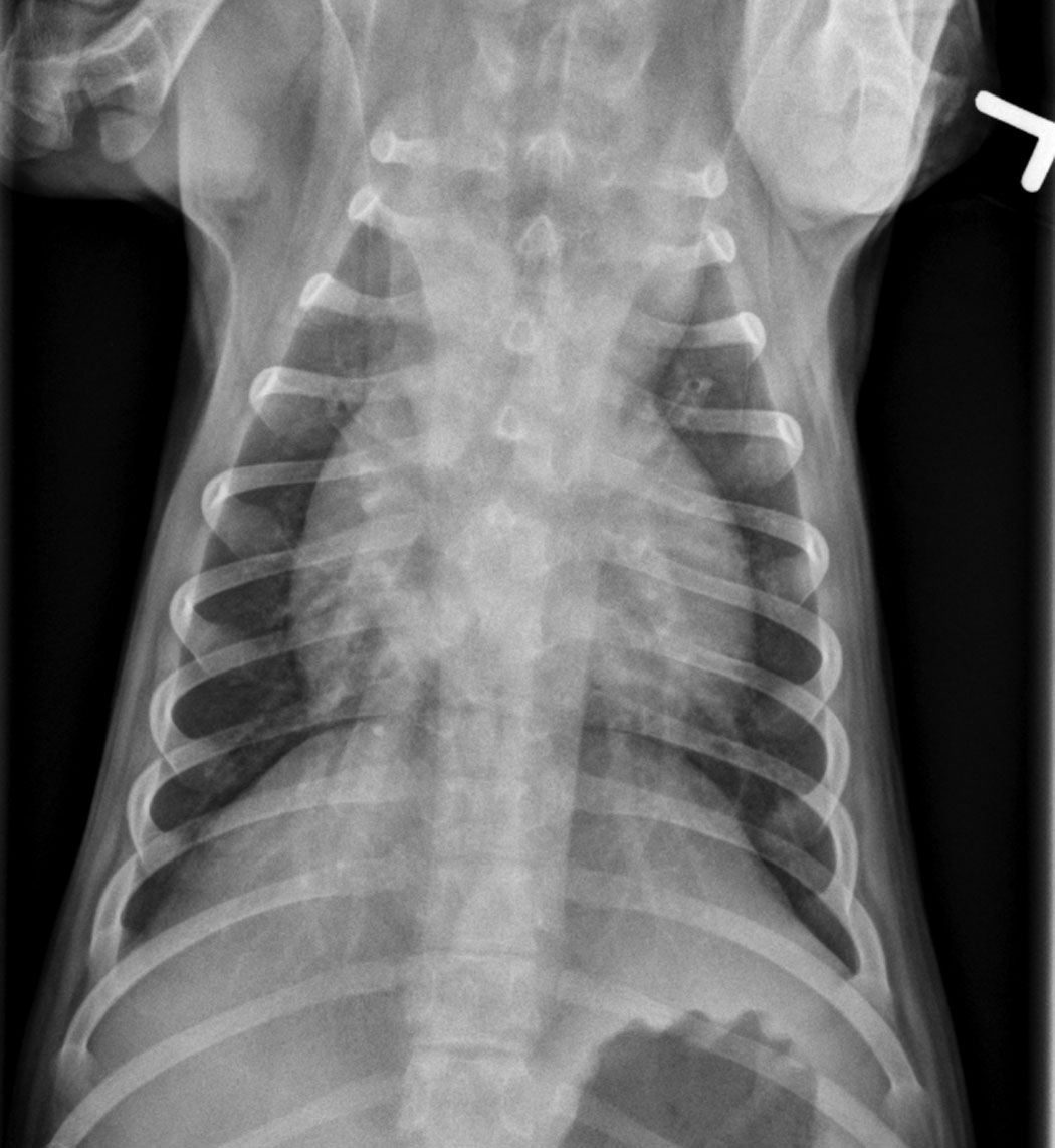
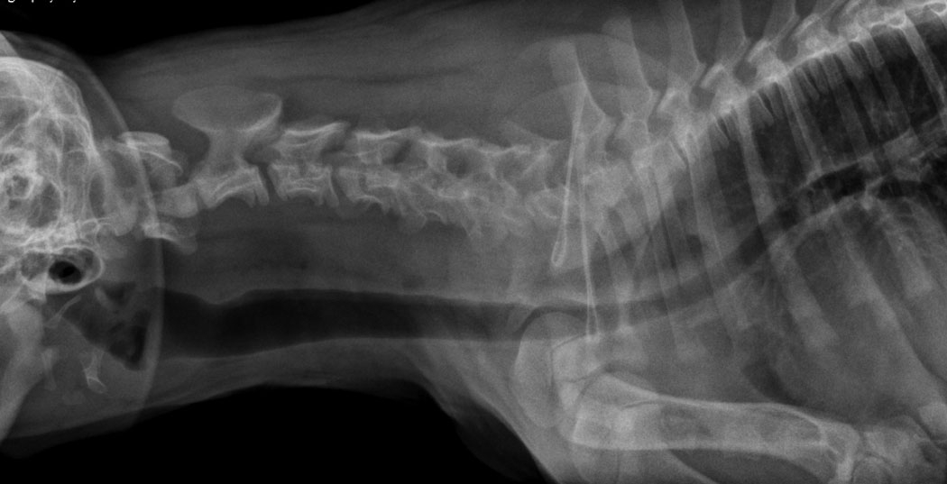
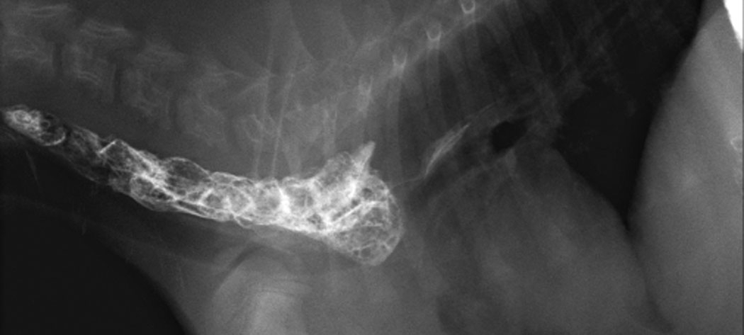
On plain radiographs, there is widening of the cranial mediastinum and soft tissue and gas opacity in the cervical esophagus. The caudal portion of the esophagus appears normal. There is no evidence of aspiration pneumonia. The trachea is displaced ventrally on the lateral projection and to the left (on midline) on the d/v.
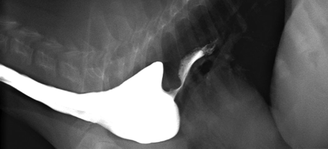
On spot film images during an esophagram, the esophagus is dilated to the level of the third rib. It narrows abruptly at this level, and very little barium and no kibble is able to pass.
Diagnosis
- Persistent right aortic arch
Discussion
The radiographic characteristics of a vascular ring anomaly include megaesophagus cranial to the restriction and leftward deviation of the trachea at the site of obstruction. The trachea and the esophagus are trapped between the aortic arch and the ligamentum arteriosum. This compression can also cause visible tracheal narrowing.
References
- Buchanan JW. Tracheal signs and associated vascular anomalies in dogs with persistent right aortic arch. J Vet Intern Med 2004;18:510-514.
- Ellison GW. Vascular ring anomalies in the dog and cat. The Compendium on Continuing Education 1980;2:693-705.
On the d/v it seems the pulmoanry veins are slightly enlarged? And the heart is deviated to the left?
The right pulmonary vein is superimposed on the caudal vena cava which might make it seem enlarged. There is always some magnification of the vasculature on this view. I think the heart is centrally located.