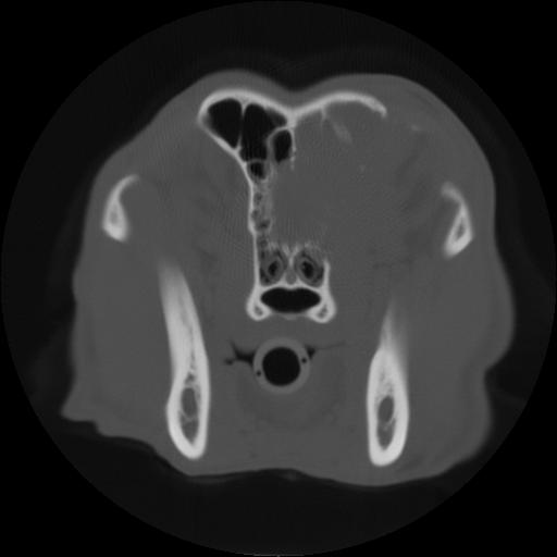
Today’s case is a 9-year-old female neutered Australian cattle dog cross with several week history of ocular discharge from the right eye, and suspected retrobulbar mass. Skull radiographs are always a challenge! Take a look and post your interpretations.
Findings
There is irregular periosteal reaction on the frontal bone on the lateral projection. There also appears to be cortical destruction of the frontal bone medial to the right mandible on the d/v projection. The right side of the cribriform plate is indistinct. There is a focal increase in soft tissue opacity in the caudal right nasal passage.
Diagnosis
Squamous cell carcinoma involving the right nasal passage, the right orbit, and the calvarium.
What about increased opacity of frontal sinus on lateral view and destruction of nasal conchae on DV view. Are they visible radiographically? If yes we should write them on radiographic findings.