Biliary mucoceles have become a common cause of extrahepatic biliary obstruction in dogs over the last two decades. The accumulation of mucinous bile in the gall bladder, cystic duct and common bile duct causes distension, and eventually rupture of the gallbladder. Many of these dogs present with … [Read more...]
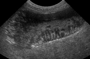
Infarction of the spleen and liver: a similar ultrasonographic pattern
Infarction of the spleen or liver is a fairly rare event. But if the blood supply is reduced, a region, or the entire organ, can become infarcted. Infarction is necrosis resulting from a reduced blood supply. The main causes of infarction of the liver and spleen are malposition (torsion) and … [Read more...]
Ultrasound of linear foreign bodies
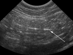
Ultrasound of linear foreign bodies can be more difficult than it seems. There are a couple of different imaging presentations depending on the size of the foreign body itself. I like to use radiographs to look for the pattern of linear foreign body, then confirm with ultrasound and look for … [Read more...]
Making the most of body position in veterinary ultrasound
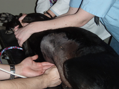
Abdominal ultrasonography is extremely useful and versatile in investigating diseases in companion animals. We as veterinarians have the advantage over human medicine; our patients are small enough that we get excellent images. When you are scanning a 150 pound Rottweiler, you experience what human … [Read more...]
Ultrasound of the adrenal glands in dogs
The adrenal glands are hard to find on ultrasound. They are less than 1 cm thick, and hide in the fat next to the aorta and caudal vena cava. Vet students and lab participants alike struggle to find these tiny organs. What can you use as landmarks? The abdominal vessels are excellent landmarks to … [Read more...]
- « Previous Page
- 1
- …
- 4
- 5
- 6
- 7
- Next Page »
Recent Comments