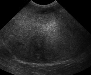
A 14 year old dog came into our hospital last week with neurological signs, and was scheduled for an MR of the brain. He came to ultrasound for an abdominal scan to investigate the cranial organomegaly that the clinican palpated when he arrived. The first image that appeared on the ultrasound screen … [Read more...]
The “A-ha” moment
One of the best things about being a radiologist is teaching others how to improve their ultrasound skills. After countless classes and labs, learning the concepts of ultrasound imaging and practicing on normal dogs, our senior students finally get into the clinic and the transducer is in their … [Read more...]
Splenic nodules
Splenic nodules or masses are extremely common findings when ultrasounding the canine abdomen. Nodules are small, circular abnormalities within the spleen that might be hyperechoic (brighter) or hypoechoic (darker) than the surrounding, normal spleen. Nodules are small (less than 4 cm diameter) and … [Read more...]
Hyperechoic fat on abdominal ultrasound – what does it mean?
Hyperechoic fat The abdominal organs are surrounded by mesentery and omentum. On radiographs, the fat stored in these structures are what allow us to see the serosal surfaces of the soft tissue density organs. Fat is hyperechoic on ultrasound, giving a fairly uniform background for the abdominal … [Read more...]
Abdominal ultrasound in canine lymphoma: expect the unexpected
Abdominal ultrasound is a routine part of staging a new lymphoma patient. The liver and spleen are often involved, as well as intra-abdominal lymph nodes. So what do we expect to see if these organs are infiltrated with lymphoma? The liver is very commonly infiltrated with neoplastic cells in … [Read more...]
- « Previous Page
- 1
- …
- 3
- 4
- 5
- 6
- 7
- Next Page »
Recent Comments