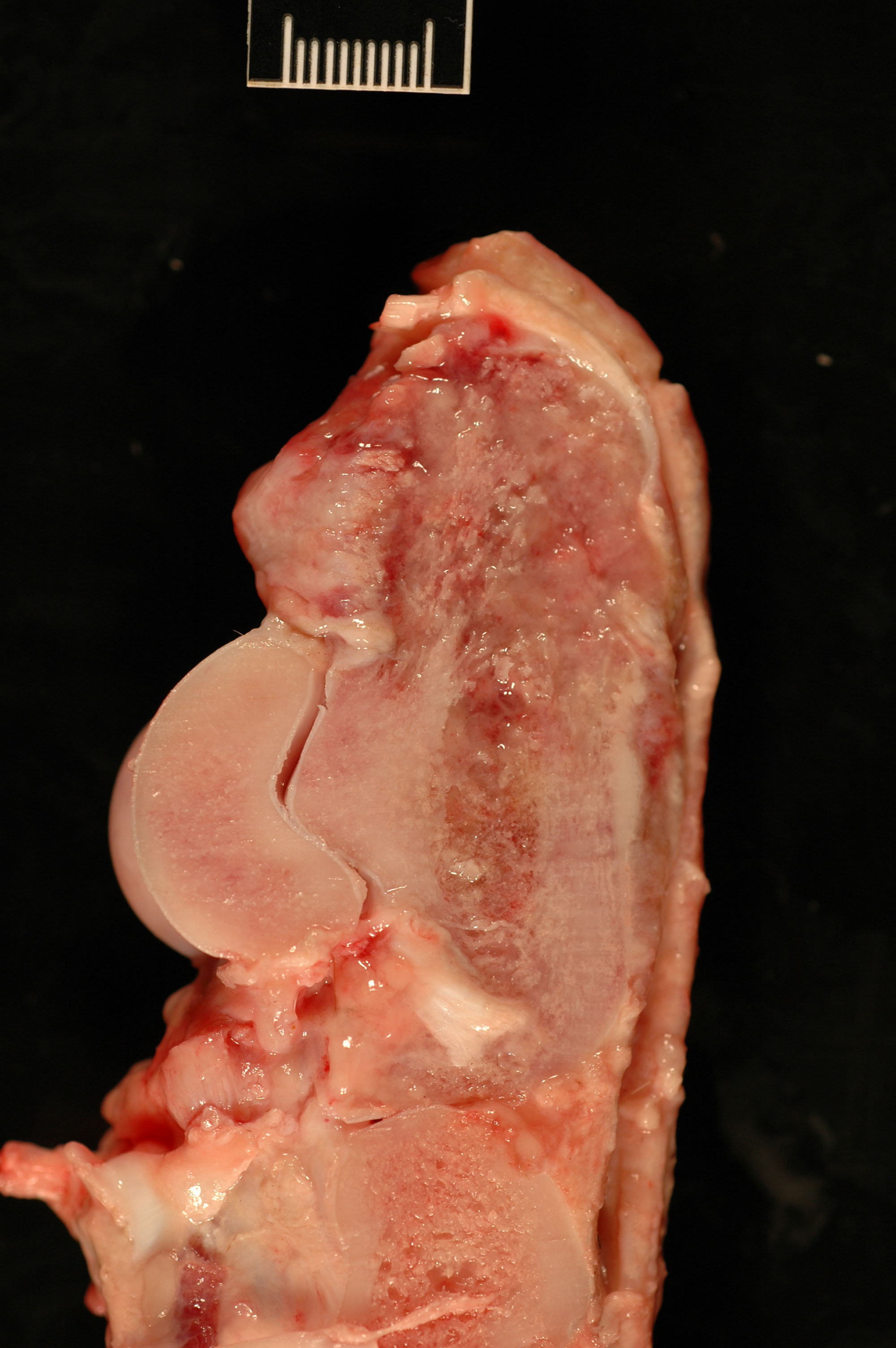
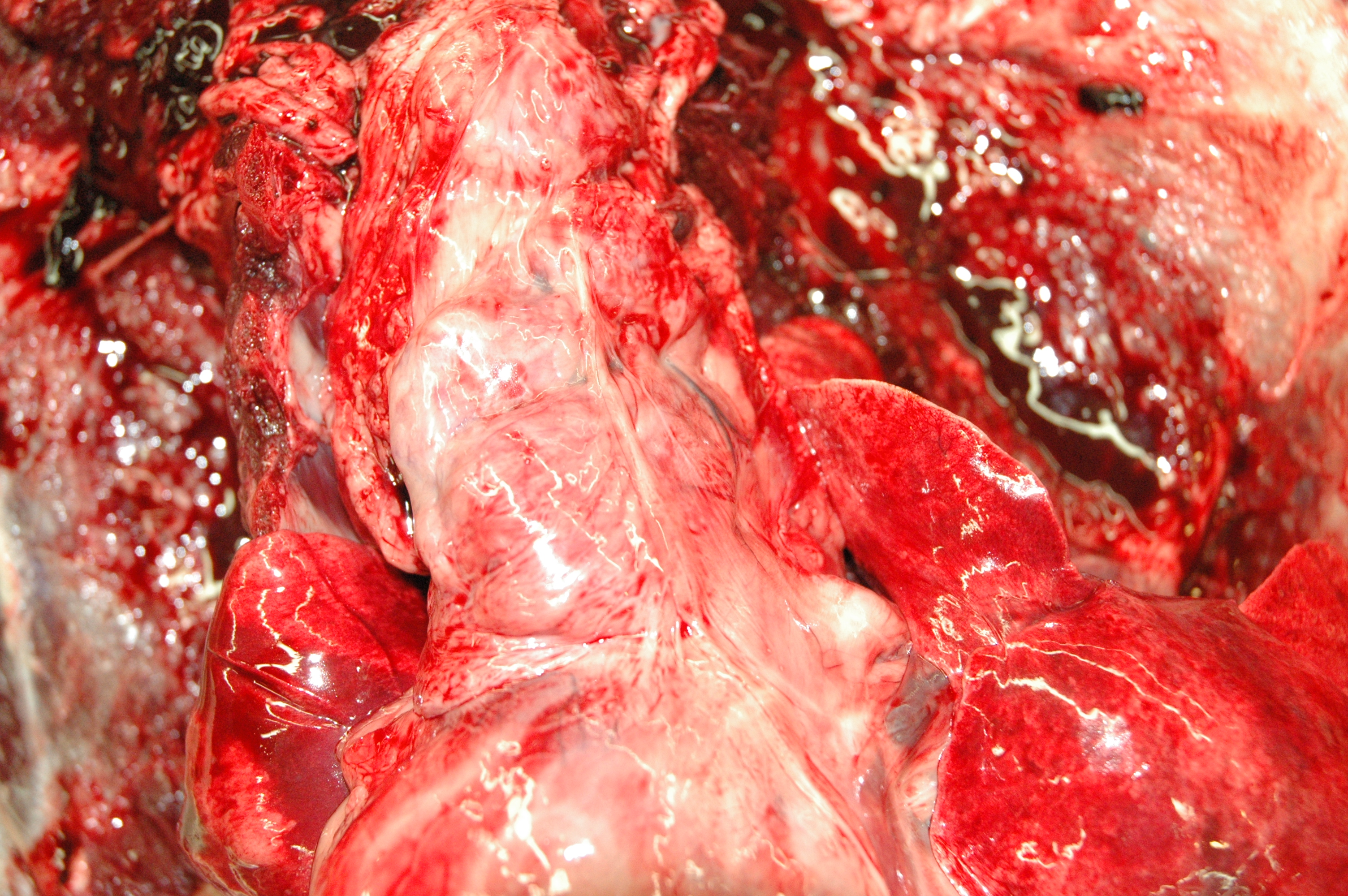
This week’s case is a 3-year-old male Rhodesian Ridgeback with swelling of the left tarsus and recent anorexia. What are your interpretations?
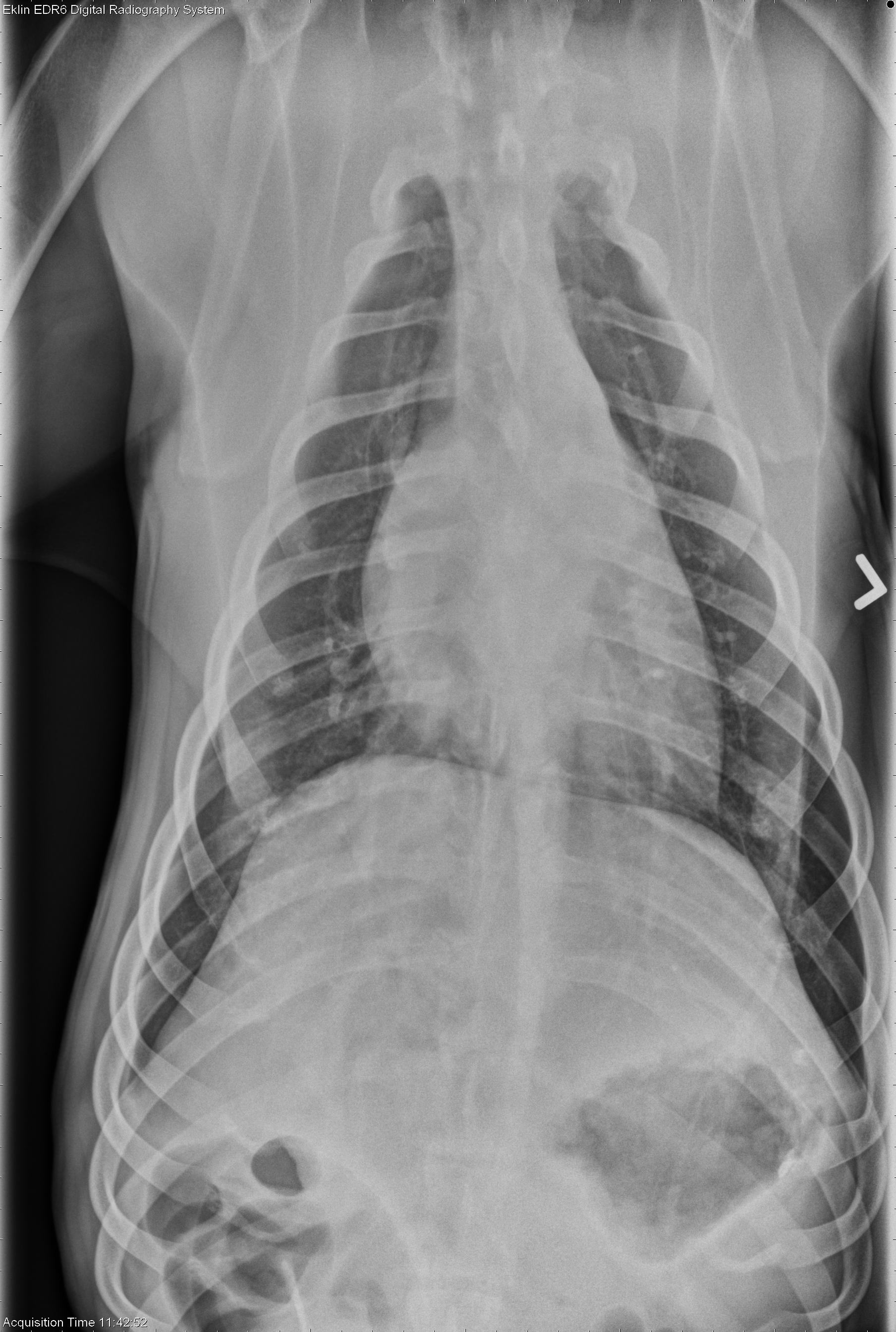
Findings
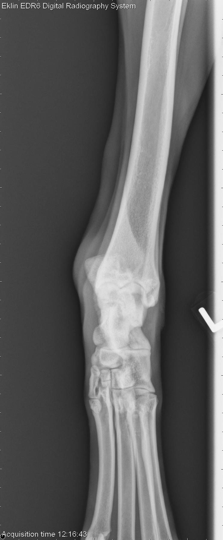
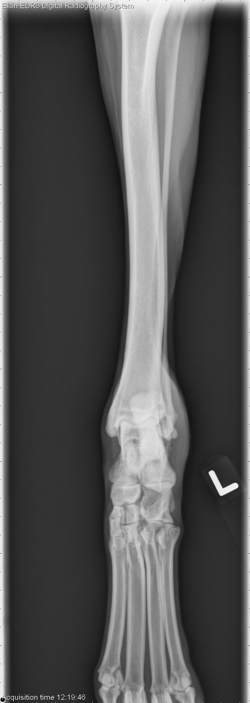
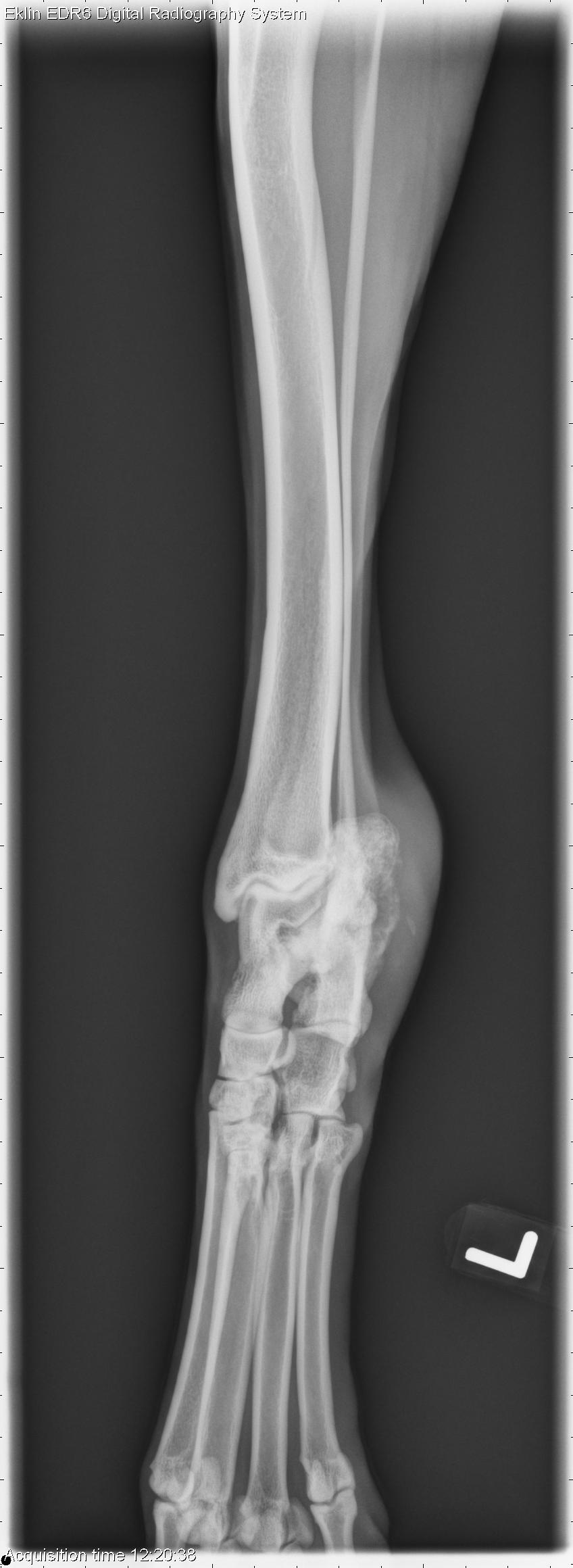
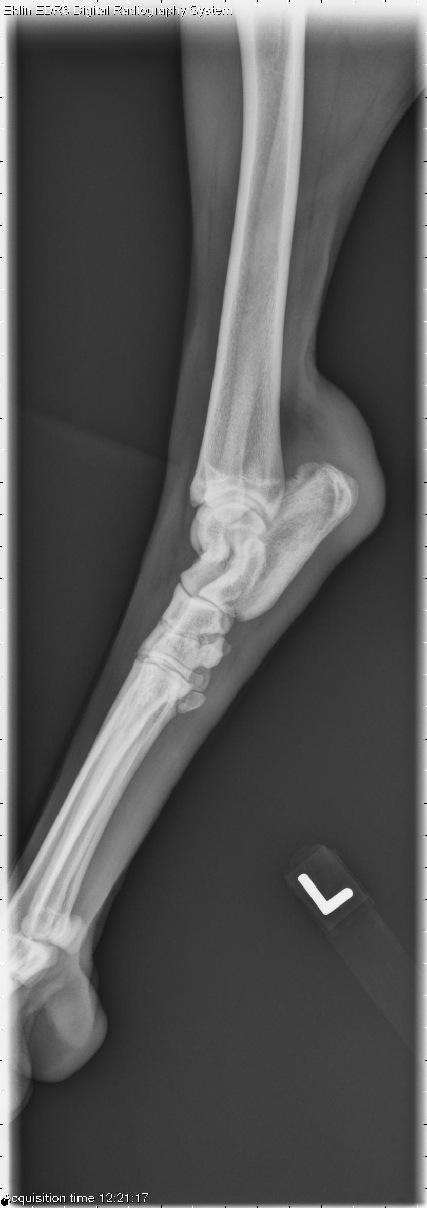
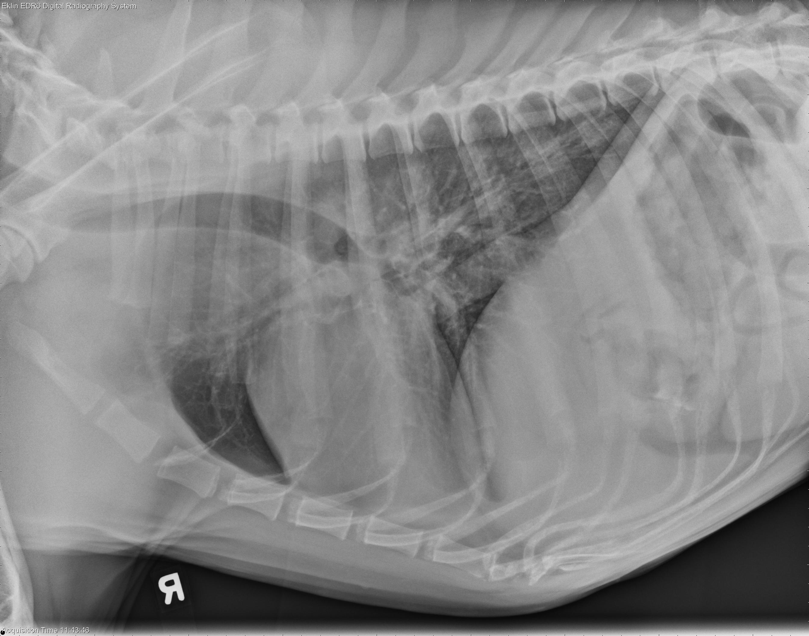
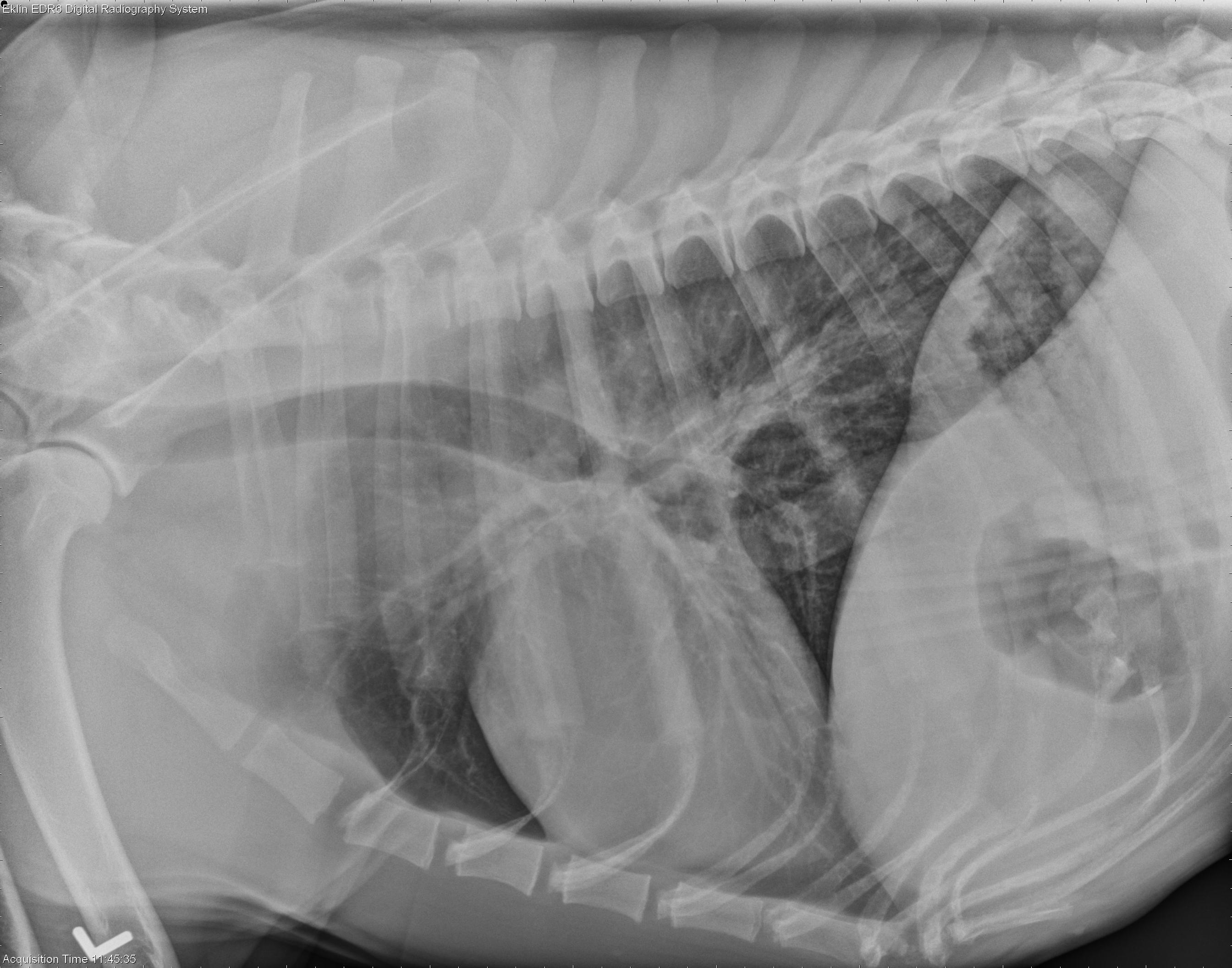
Tarsus – There is an aggressive bone lesion of the lateral and mid to proximal aspect of the left calcaneus. There is cortical destruction and marked, irregular periosteal proliferation. There is associated extracapsular soft tissue swelling at this site. There is a small, elongated mineral fragment within this soft tissue. the calcaneal tendon is thickened and irregular.Thorax – There is increased opacity of the cranial mediastinum on the lateral projection, with dorsal tracheal deviation. The cranial mediastinum is mildly widened on the d/v projection. There is also ill-defined increase in soft tissue opacity dorsal to the carina. In addition, there is a mild, diffuse interstitial pulmonary pattern. The cardiovascular structures are within normal limits. There are small mineral opacities within the stomach.
Differential Diagnosis
aggressive bone lesion and lymphadenopathy – systemic mycosis, systemic bacterial infection, neoplasia (less likely)Diagnosis
Systemic aspergillosisDiscussion
Aggressive bone lesions such as these are most often due to osteomyelitis or neoplasia. The finding of enlarged thoracic lymph nodes makes an infectious process most likely. Fungal infection typically causes marked enlargement of thoracic lymph nodes.Abdominal ultrasound was indicated but not performed in this case. Necropsy revealed a renal mass and a diffusely enlarged, mottled spleen as well as lymphadenopathy. Schultz RM, Johnson EG, Wisner ER, et al. Clinicopathologic and diagnostic imaging characteristics of systemic aspergillosis in 30 dogs. J Vet Intern Med 2008;22:851-859.
Additional images