Today’s case is an 8-year-old male neutered Bichon Frise adopted from a rescue organization. Presents for coughing. What are your findings?
Findings
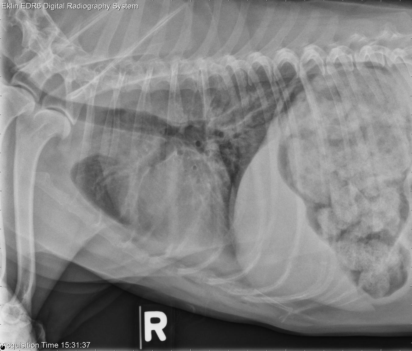
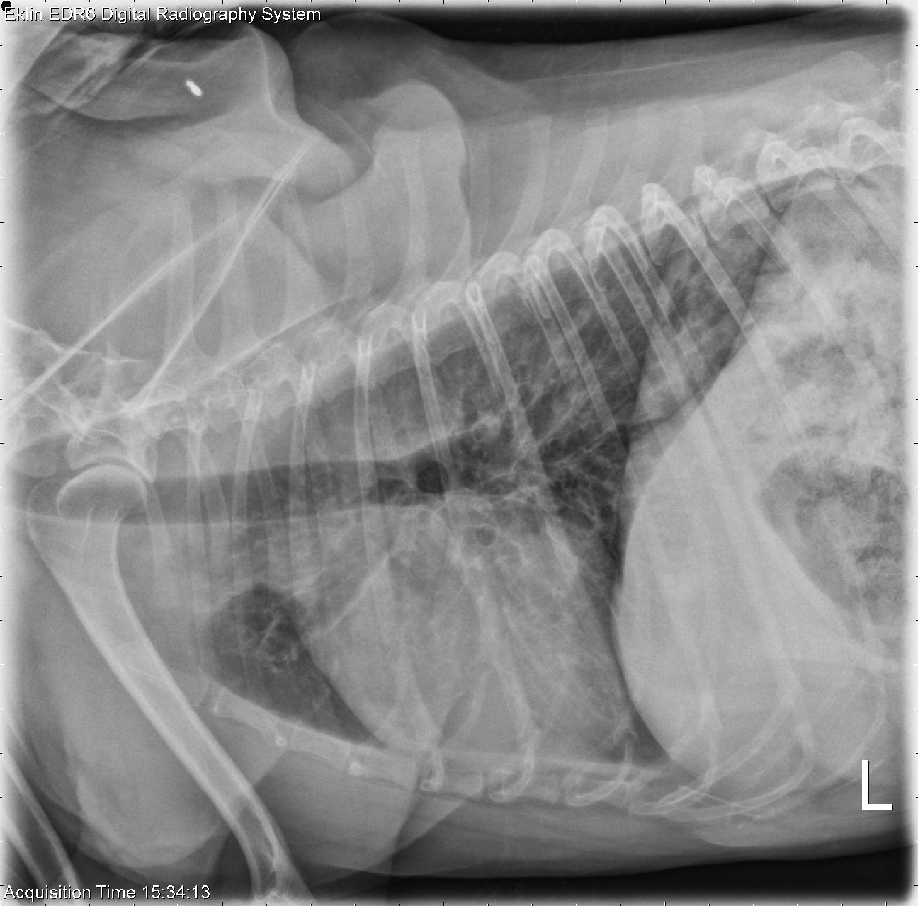
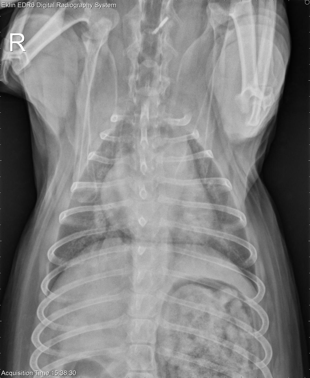
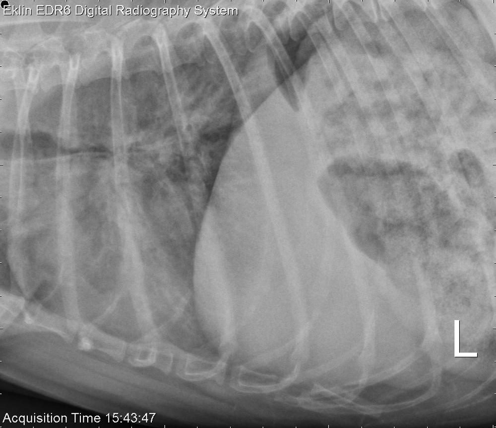
There is hypoinflation of the lungs on all projections. The bronchi have irregularly thickened walls, causing a bronchial pattern. The airways do not taper in the periphery of the lung and appear widened. There is a diffuse interstitial pattern in the remainder of the lung. There is an ill-defined alveolar opacity on the left lateral projection superimposed on the 8th intercostal space. On the final lateral radiograph taken during expiration, there is complete collapse of the carina and mainstem bronchi. The cardiovascular structures, pleural space, and mediastinum appear normal.
Differential Diagnosis
- Chronic inflammatory and/or noninflammatory lower airway disease with bronchiectasis
- Diffuse pulmonary inflammation or fibrosis
- Possible inflammatory focus or pulmonary nodule
- Lower airway collapse
Diagnosis
Chronic bronchitis and bronchiectasis with lower airway collapse.
The focal alveolar pattern resolved on subsequent thoracic radiographs and was likely inflammatory.