• Home
  • About
  • Articles
  • Books
  • Contact

Veterinary Radiology

Teaching and learning about veterinary diagnostic imaging.

  • Modality
    • Radiographs
    • Ultrasound
    • CT/MRI
  • Region
    • Thorax
    • Abdomen
    • Musculoskeletal
    • Neurologic
  • Species
    • Canine
    • Feline
    • Equine
    • Exotic

15 year old DSH

April 6, 2015 By Allison Zwingenberger

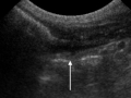
This week’s case is a 15 year old domestic shorthair referred for chronic obstipation and weight loss.

R LAT Abdomen
VD Abdomen

Show findings...

Findings

There is dilated bowel occupying the majority of the abdomen, giving it a distended appearance. The dilated loops are filled with fluid and mineral opacity material. The loops could be colon; however, they seem more extensive than the normal amount of large intestine. There is a small amount of gas in the terminal colon and rectum, which is not filled with fecal material.

Show additional images...

Additional images

R LAT barium enema
VD barium enema

A barium enema confirmed that the dilated bowel was not colon.

Ultrasound-annotated

There was limited visibility in the abdomen. The dilated segment of bowel tapered into a narrowed area (arrow) with some loss of wall layering and hypoechoic echogenicity. At surgery, the adenocarcinoma appeared as a fibrotic strictured area.

Show differential diagnosis…

Differential Diagnosis

  • Chronic partial small intestinal obstruction
  • Megacolon

Show diagnosis…

Diagnosis

Stricture due to adenocarcinoma of the ileum.

Show discussion…

Discussion

A barium enema was performed (see additional images), which confirmed that the dilated bowel was not large intestine in origin. The mineral material in the small intestine oral to the obstruction is characteristic of a “gravel sign”, or accumulation of particulate matter that is too large to pass through the intestinal lumen at the site of obstruction.
Ultrasound was performed; however, visibility was limited due to the large amount of attenuating material in the colon.

Case originally posted on February 26, 2007

Filed Under: Abdomen, Case of the Day, Feline, Radiographs

Comments

  1. Vidalini says

    April 14, 2015 at 7:21 am

    Massive Megacolon!

  2. Vidalini says

    April 14, 2015 at 7:24 am

    Ups!! Guess I jumped into conclusions to soon! Amazing case!

  • Bloglovin
  • Facebook
  • RSS
  • Twitter
AtlasCover787x1024 amazon-availablenow

Veterinary Radiology News

Sign up for alerts about new cases and newsletters.

We will respect your privacy.

Archives

Recent Comments

Tags

abscess adenocarcinoma bronchiectasis carcinoma cardiomyopathy coccidioidomycosis discospondylitis elbow dysplasia eosinophilic bronchopneumopathy erosive polyarthritis feline infectious peritonitis FIP foreign body fracture gastric dilation gastric foreign body gastrointestinal GDV heart failure hemangiosarcoma hiatal hernia histiocytic sarcoma hypertrophic cardiomyopathy intestinal foreign body linear foreign body lymphoma megaesophagus osteochondrosis osteomyelitis osteosarcoma patent ductus arteriosus PDA pericardial effusion pneumonia pneumothorax polyarthritis PPDH pulmonary pulmonary abscess pulmonary adenocarcinoma pulmonary carcinoma sarcoma thymoma tracheal collapse vascular ring anomaly

Copyright © 2025 · Metro Pro Theme On Genesis Framework · WordPress · Log in