• Home
  • About
  • Articles
  • Books
  • Contact

Veterinary Radiology

Teaching and learning about veterinary diagnostic imaging.

  • Modality
    • Radiographs
    • Ultrasound
    • CT/MRI
  • Region
    • Thorax
    • Abdomen
    • Musculoskeletal
    • Neurologic
  • Species
    • Canine
    • Feline
    • Equine
    • Exotic

Renal enlargement in veterinary radiology

February 23, 2007 By Allison Zwingenberger

Renal disease is extremely common in dogs and cats. In veterinary diagnostic imaging, we use radiographs, ultrasound and sometimes special studies to determine if one or both kidneys are affected. Radiographs are often a good place to start to determine renal size and detect calculi.

Left renal enlargementLeft renal enlargement

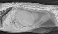
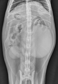
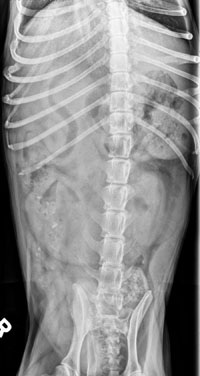
The normal reference range sizes for kidneys are 2.5-3.5 x the length of L2 for dogs, and 2-3 x the length of L2 for cats. It’s measured best on the v/d projection where the kidneys remain in a more consistent position.

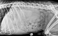
The left kidney usually sits caudal to the right kidney, with the cranial pole at L2 in the dog and cat. It resides in the retroperitoneal space, dorsal and lateral to the descending colon. If the left kidney enlarges, it tends to displace the descending colon medially and ventrally.

The right kidney is located dorsal to the descending duodenum and the ascending colon. Enlargement of the right kidney can cause medial and ventral displacement of the descending duodenum, and medial and ventral displacement of the ascending colon.

Right kidney enlargementRight kidney enlargement

Filed Under: Uncategorized

  • Bloglovin
  • Facebook
  • RSS
  • Twitter
AtlasCover787x1024 amazon-availablenow

Veterinary Radiology News

Sign up for alerts about new cases and newsletters.

We will respect your privacy.

Archives

Recent Comments

Tags

abscess adenocarcinoma bronchiectasis carcinoma cardiomyopathy coccidioidomycosis discospondylitis elbow dysplasia eosinophilic bronchopneumopathy erosive polyarthritis feline infectious peritonitis FIP foreign body fracture gastric dilation gastric foreign body gastrointestinal GDV heart failure hemangiosarcoma hiatal hernia histiocytic sarcoma hypertrophic cardiomyopathy intestinal foreign body linear foreign body lymphoma megaesophagus osteochondrosis osteomyelitis osteosarcoma patent ductus arteriosus PDA pericardial effusion pneumonia pneumothorax polyarthritis PPDH pulmonary pulmonary abscess pulmonary adenocarcinoma pulmonary carcinoma sarcoma thymoma tracheal collapse vascular ring anomaly

Copyright © 2025 · Metro Pro Theme On Genesis Framework · WordPress · Log in