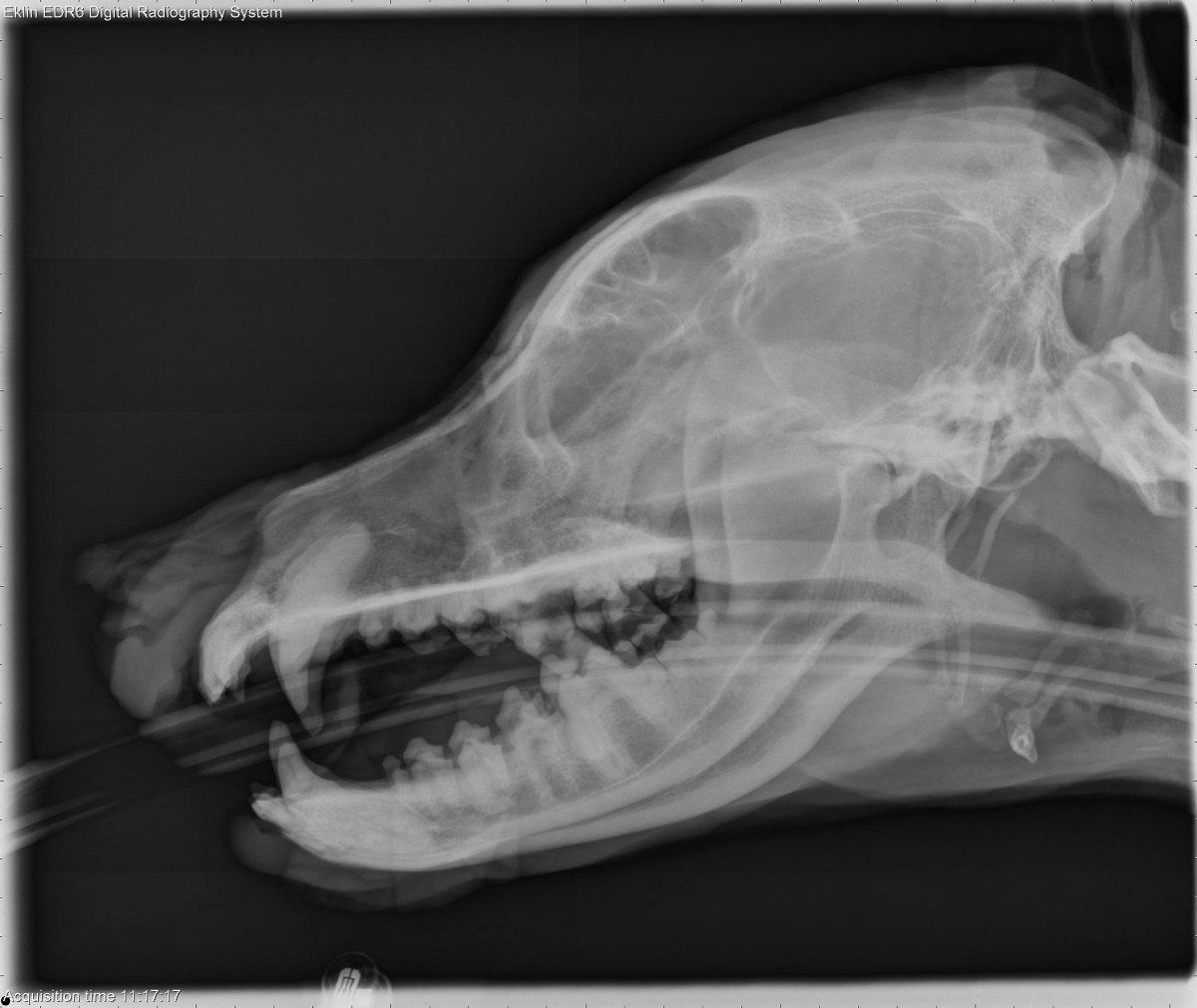
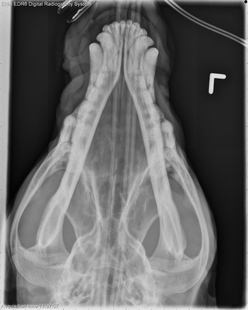
Today’s case is an 11-year-old male mixed breed dog with respiratory distress. We don’t see skull radiographs very often. Can you spot the abnormalities?
Teaching and learning about veterinary diagnostic imaging.
Today’s case is an 11-year-old male mixed breed dog with respiratory distress. We don’t see skull radiographs very often. Can you spot the abnormalities?