Today’s case is a 1.5 year old female neutered Boxer with one-month history of a cough and dermal masses. What are your findings?
Findings
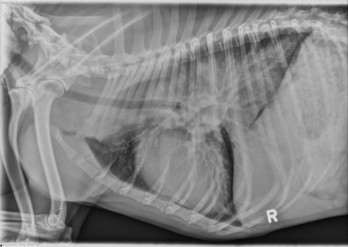
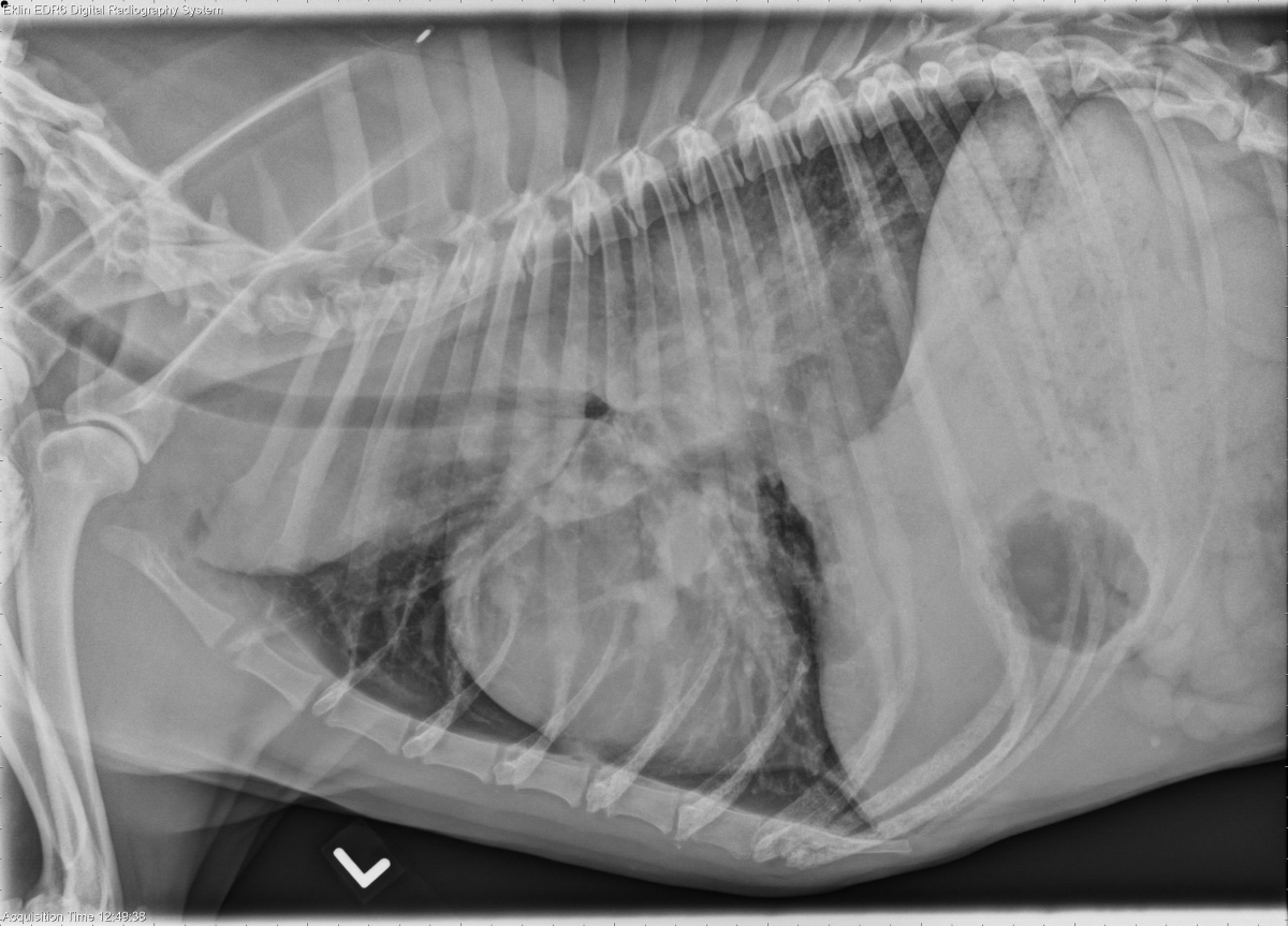
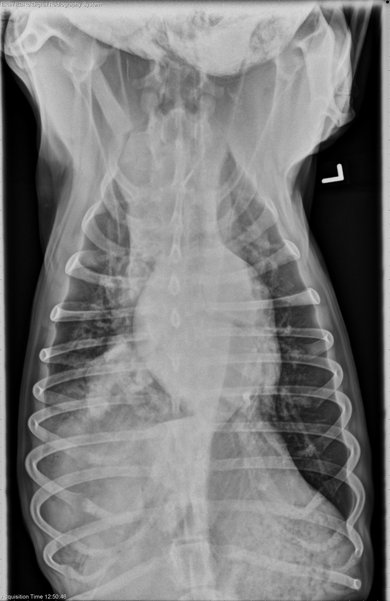
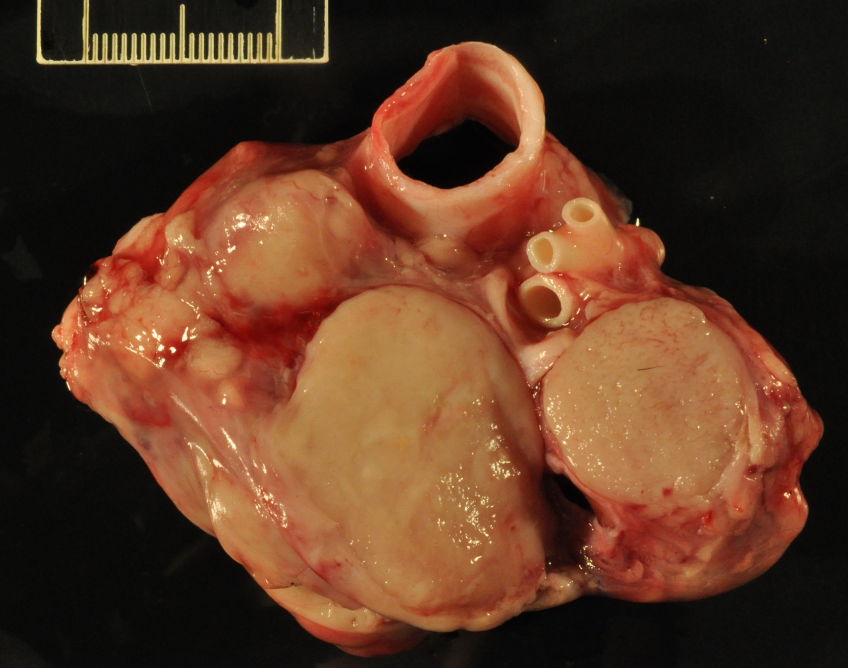
There is a large mass in the right caudal lung lobe causing an alveolar pattern and lobar sign. Additional masses are present in the right middle lung lobe. There is a diffuse bronchointerstitial pattern. The cranial mediastinum is widened. The mainstem bronchi are compressed, and caudal mainstem bronchi are displaced ventrally and laterally. There are soft tissue masses in the region of the carina. A small amount of air is present in the pleural space on the right lateral projection.
Differential Diagnosis
Pulmonary masses and mediastinal and tracheobronchial lymphadenopathy are likely secondary to disseminated neoplasia, such as lymphoma or histiocytic sarcoma, or granulomatous disease. The bronchointerstitial pattern may be due to additional infiltrate. Mild pneumothorax may be secondary to cough or iatrogenic.
Diagnosis
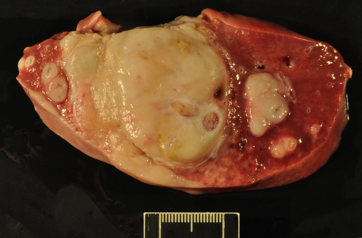
The dermal nodules, mediastinal mass, and pulmonary masses were due to large granular T-cell lymphoma.
Discussion
Boxers have a breed disposition to T-cell lymphoma.