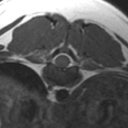
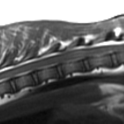
This week’s case is an 11-month-old Rottweiler. It is a congenital neurological problem that you don’t see every day, but it is a real classic. Take a look through the axial and sagittal MRI series below.
Findings
There is a mass at the level of T11, which is approximately half the length of a vertebral body. At the cranial aspect, the mass is broad based peripherally and extends into or compresses the spinal cord. This suggests an intradural-extramedullary lesion. More caudally, the mass appears intramedullary. The mass is hypointense on T2, slightly hyperintense on T1, and is mildly contrast enhancing. There is hyperinteinsity of the spinal cord on T2 cranial and caudal to the lesion.Differential Diagnosis
Lymphosarcoma Hyperintense spinal cord on T2- gliosis
- edema
- syrigohydromyelia
Diagnosis
NephroblastomaDiscussion
Nephroblastomas are remainders of renal tissue. They occur between T10-L2 and are intradural-extramedullary. This may be difficult to determine on myelography and MR if the tumor is subpial, and it may appear intradural. MR is better than myelography at distinguishing between these locations. Signs of an intradural-extramedullary tumor include widening of the subarachnoid space and outward displacement of epidural fat, which were not visible in this case. Another reported sign in people is a T1 hypointense band between the mass and the spinal cord which may represent tumor capsule and trapped CSF.On MR nephroblastomas arise peripherally and compress the cord. They are lobular and may infiltrate. This mass had infiltrated the cord at surgery.
Intradural-extramedullary masses in the spinal cord of dogs have been reported as nephroblastoma, ependymoma, medulloepithelioma, neuroepithelioma, embryonal nephroma, embryonal adenosarcoma, renal adenocarcinoma and Wilms tumor. In Jubb and Kennedy, these are presumed to all arise from embryonal renal tissue, as supported by immunohistochemical staining.