Today’s case is a 12 year old male neutered English Cocker Spaniel with a soft, non-productive cough. Post your interpretations of the thoracic radiographs in the comments section.
Findings
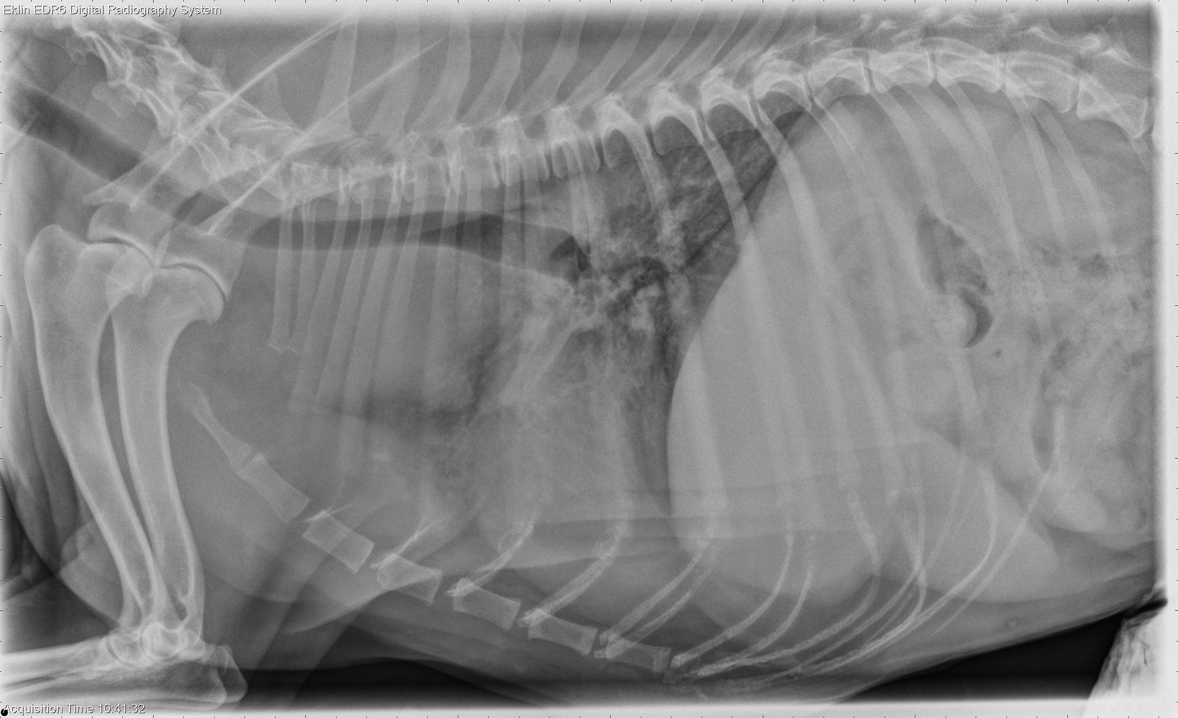
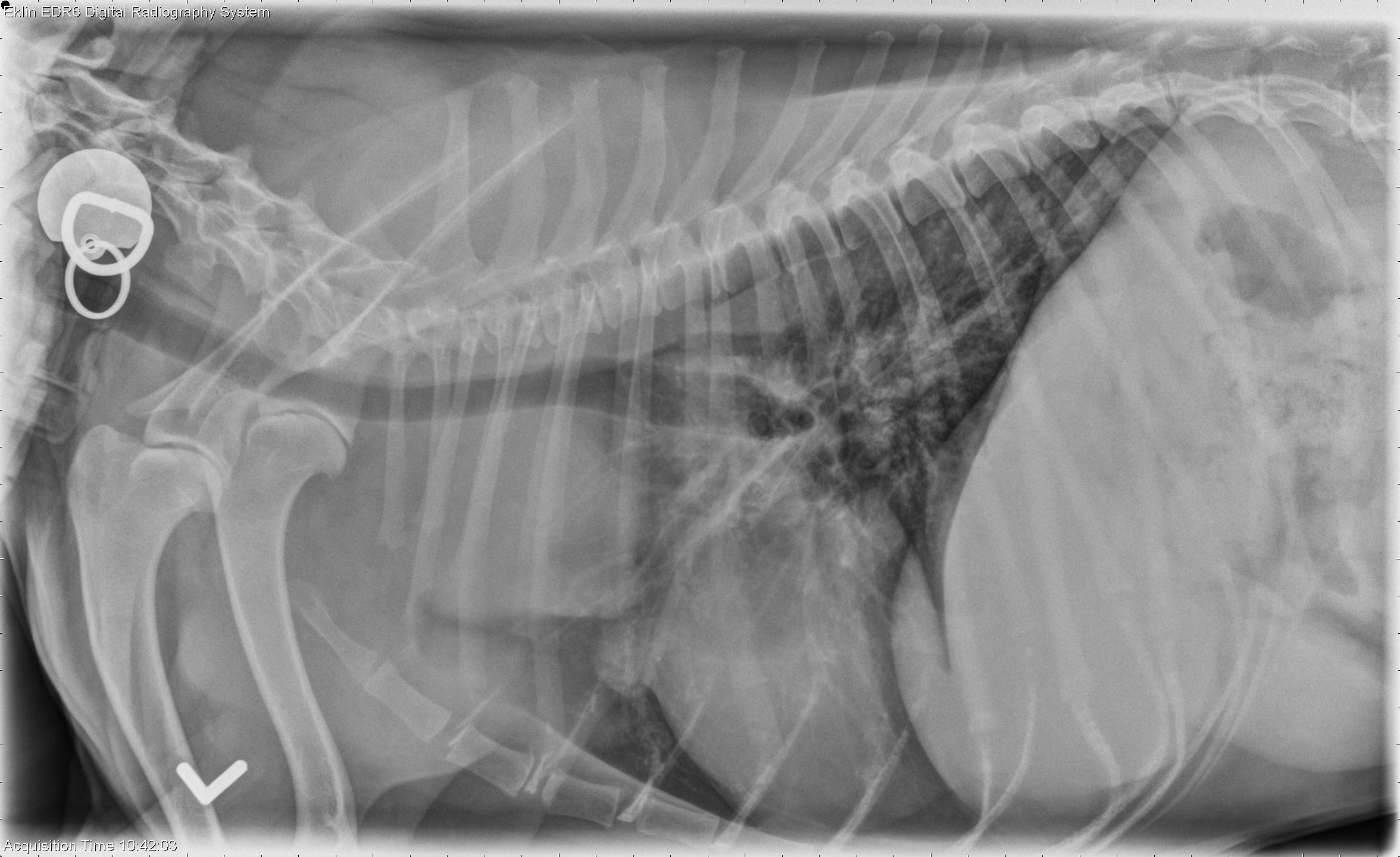
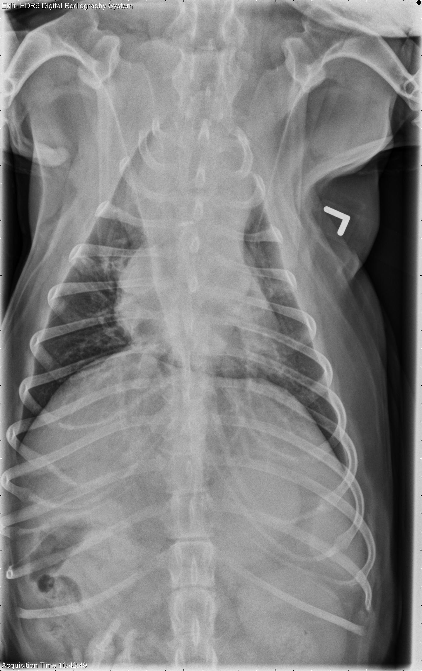
There are multiple mediastinal masses visible. There is marked enlargement of the sternal lymph node and enlargement of the hilar lymph node. There is a large soft tissue mediastinal mass that is displacing the trachea dorsally. A round soft tissue opacity is present cranial to the heart on both lateral projections which is likely in the cranial mediastinum since the caudal border of it silhouettes with the heart. The cranial mediastinum is markedly widened on the d/v projection.The liver extends caudal to the costal arch on both lateral projections and has a rounded caudal margin. The cardiovascular structures appear within normal limits.
Differential Diagnosis
- Sternal and mediastinal lymphadenopathy indicates lymphosarcoma.
- Hepatomegaly may be due to lymphosarcoma, inflammatory disease or steroid hepatopathy.