Everyone has had those frustrating cases where you think your patient might have ingested some sort of foreign body, and is persistently vomiting. You’ve done repeat radiographs, and are debating between an upper GI and ultrasound. Could there be something radiolucent in the GI tract that you could find quickly with ultrasound?
There are pros and cons to using ultrasound for foreign body hunts. It is faster than an upper GI, and provides more detail about the GI tract integrity and sometimes about the foreign body itself. The main drawback is not being able to run the small intestine from end to end, and worrying if you missed the lesion.
As is often the case, ultrasound artifacts can actually help you to identify a foreign body in the GI tract. Just like we use distal acoustic enhancement to identify fluid-filled structures, foreign objects often have a signature of their own. 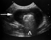 Empty or fluid filled bowel loops are easy to identify and determine that there is nothing in the lumen. Those with gas are more difficult, as the ultrasound waves reverberate from the gas to the transducer over and over, obscuring your view of the lumen. But this grey, speckled “dirty shadowing” tells you that the bowel loop is filled with gas, not a foreign body (image 1, ^). Conversely, foreign bodies are made of solid material most of the time, and tend to absorb sound rather than reflect it. Within the lumen, they actually attenuate the ultrasound waves, leaving a black, or “clean shadow” (image 2, *).
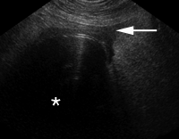
Other clues that will lead you to the site of obstruction are dilated, fluid filled loops on the oral side of the obstruction, and hyperechoic mesentery if there is any leakage or inflammation around the obstructed site. Peritoneal effusion is a sign of impending or existing bowel rupture, and a sample is useful to see if emergency surgery is required (image 2, arrow). Hopefully you will not see free gas in the uppermost portion of the abdomen that means bowel rupture has already occurred.
Older dogs and cats are a particular worry because they tend to be more sensible about eating foreign material. In these types of patients, I make sure to evaluate the bowel for any masses that could be causing a partial obstruction. These generally appear as thickening and loss of layering of the bowel wall (image 1, arrow). Image 1 is an example of lymphoma in a cat, and image 2 is a small intestinal foreign body.
Ultrasonographic signs of small intestinal obstruction:
Recent Comments