Today’s case is an 8 year old female neutered Siamese cat with a 2-week history of increased respiratory effort and wheezing. What are your findings?
Findings
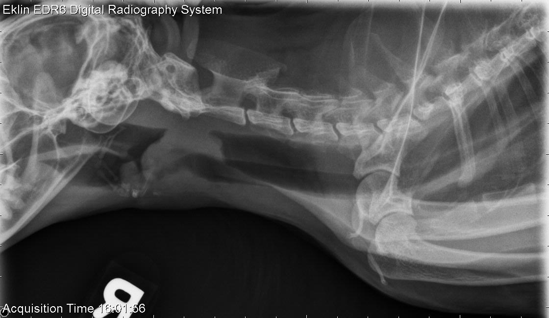
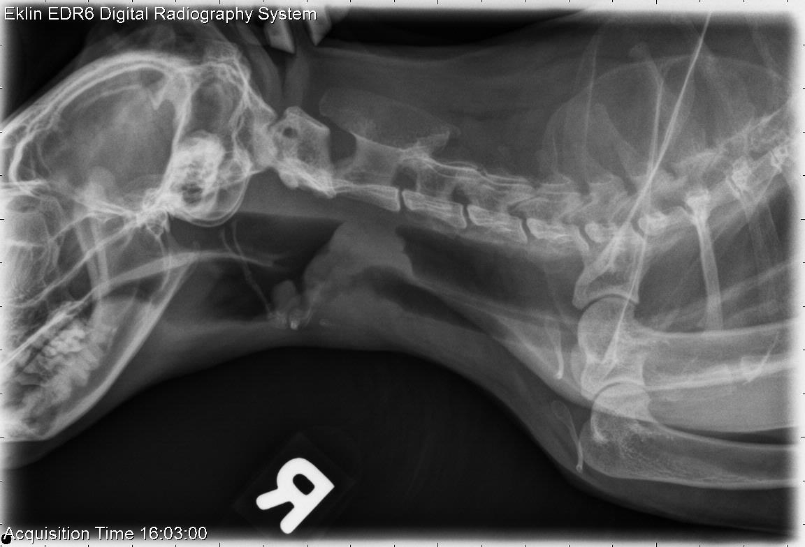
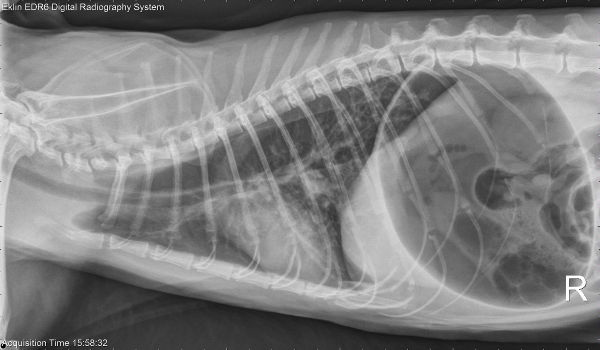
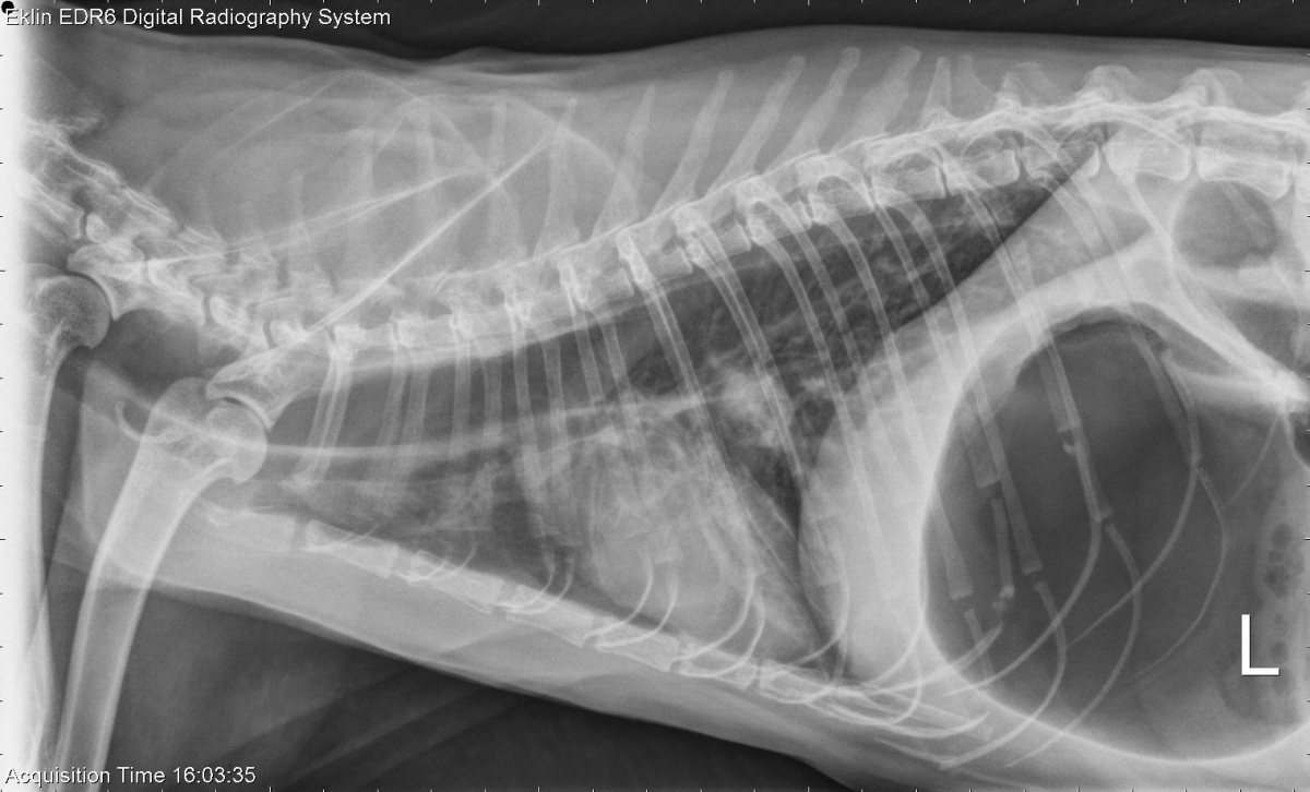
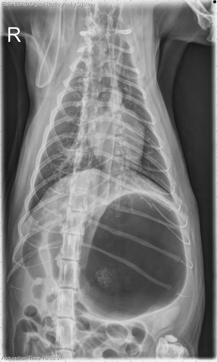
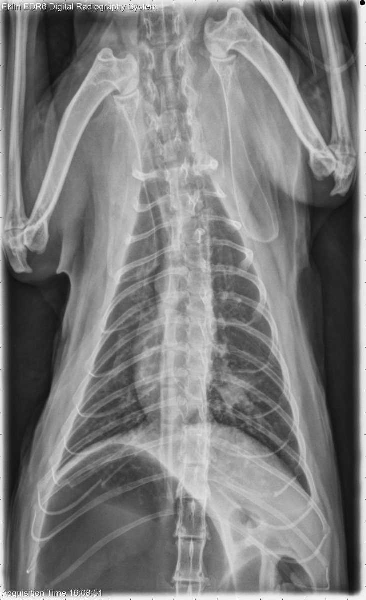
On radiographs of the neck, there is dilation of the nasopharynx and oropharynx with mild retraction of the hyoid apparatus. There is increased soft tissue opacity in the larynx with irregular margins, extending into the trachea. There is marked gas dilation of the esophagus and stomach. The pulmonary parenchyma has a diffuse bronchointerstitial pattern, and there is a focal nodule with ill-defined borders in the right caudal lung lobe. This is best seen on the ventrodorsal projection which is displayed with left and right reversed. The left 10th costal cartilage is fractured.
Differential Diagnosis
- Laryngeal obstruction, likely due to a mass such as lymphoma or carcinoma
- Secondary costal cartilage fracture and aerophagia
- Chronic lower airway disease which may have infectious and noninfectious inflammatory components
- Pulmonary nodule – granuloma, neoplasia
Diagnosis
- Lymphoma (intermediate cell size) with histiocytic inflammation
- Ultrasound guided aspirates of the laryngeal mass and a mass in the ileum were obtained for cytology