This week’s case is a 9-month-old female neutered Labrador Retriever with 2-month history of left thoracic limb lameness and 2-day history of left pelvic limb lameness. How many lesions can you find?
Findings
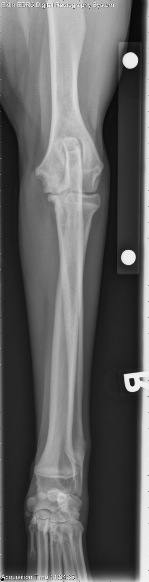
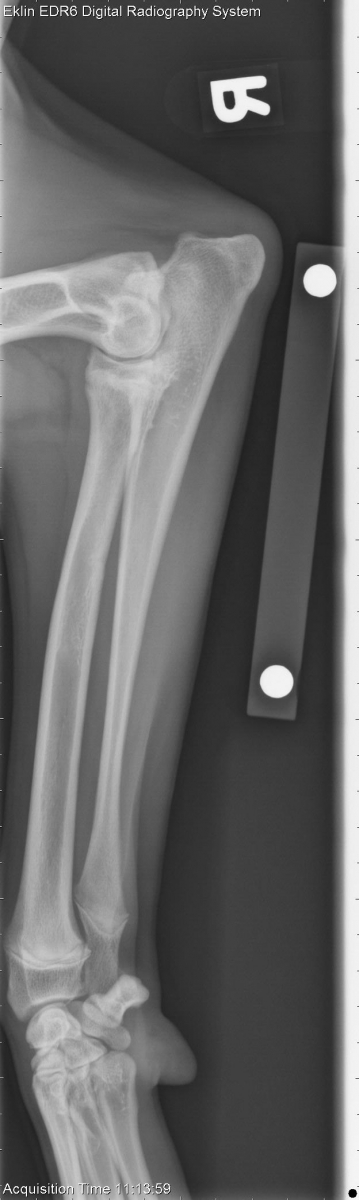
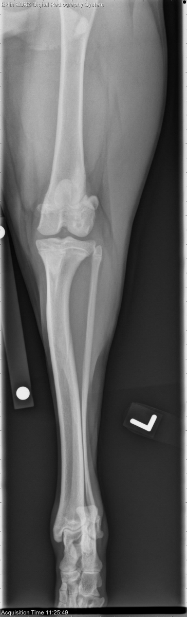
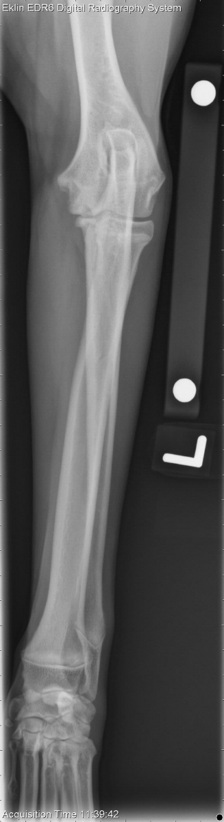
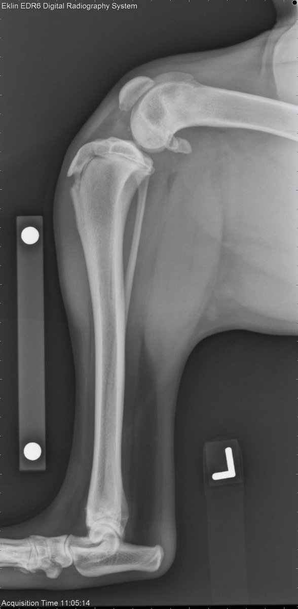
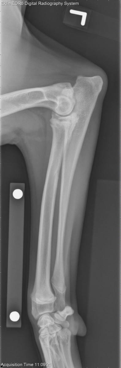
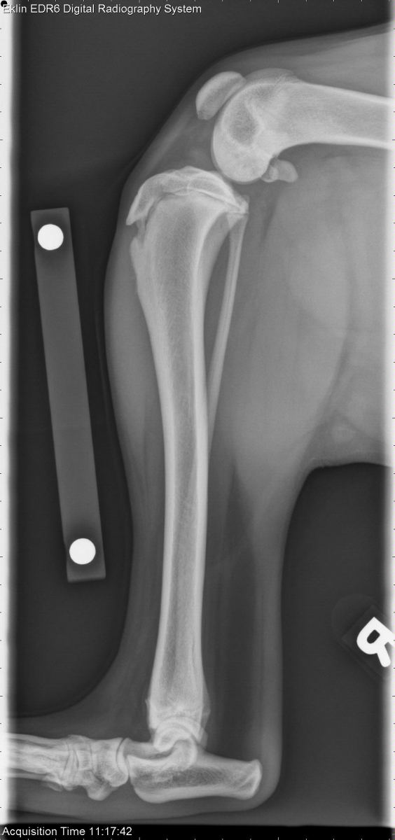
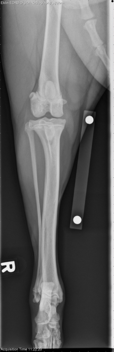
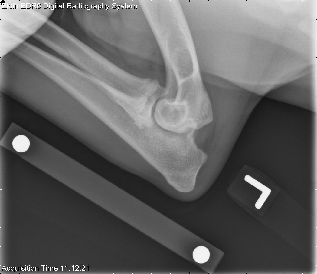
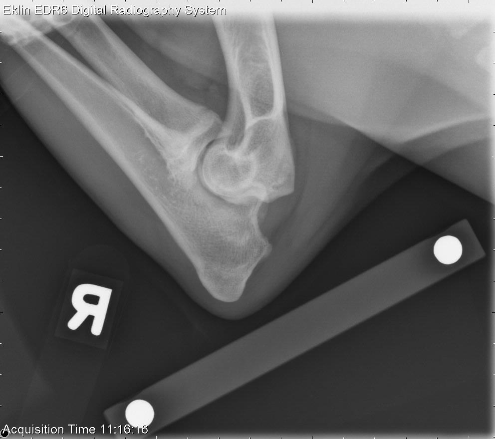
Elbows: There is incongruity of both elbows with widening of the humeroradial joint. The medial coronoid processes of the ulna are indistinct. There is osteophyte formation on the cranial radius and medial radius and ulna, as well as the non-articular portion of the anconeal process. Subchondral bone defects with surrounding sclerosis are present in both the left and right humeral condyles.Stifles: There is moderate intracapsular effusion in the left stifle and mild effusion in the right stifle. A small fragment is visible cranial to the femoral condyles on the lateral projection of the left stifle. On the caudocranial projection, there is a round radiolucency in the lateral condyle of the left stifle with a sclerotic rim. The right lateral femoral condyle is irregular in this region with ill-defined borders.
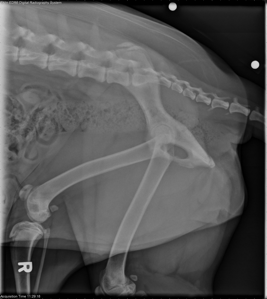
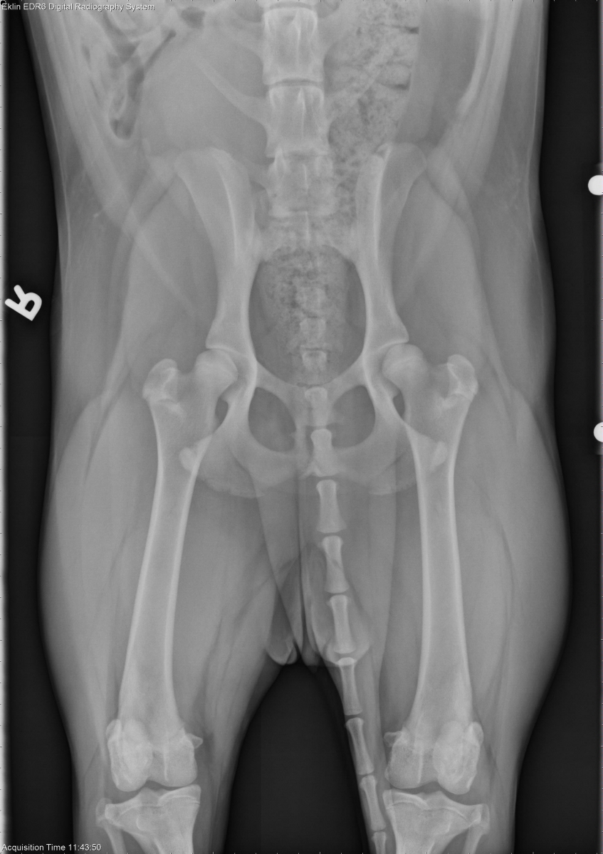
Pelvis: The coxofemoral joints are congruent and there is good coverage of the femoral head by the acetabula. No degenerative disease is noted.
Diagnosis
- Bilateral fragmented medial coronoid process of the ulna
- Bilateral osteochondrosis of the medial humeral condyle
- Probable left cranial cruciate ligament avulsion, right cranial cruciate partial/full rupture
- Suspected bilateral femoral osteochondrosis