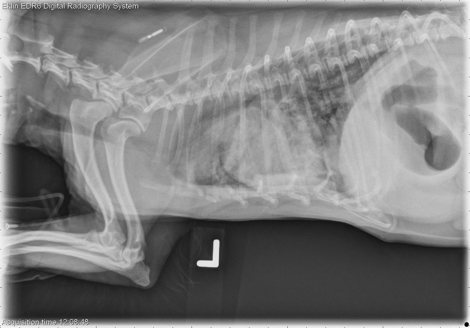
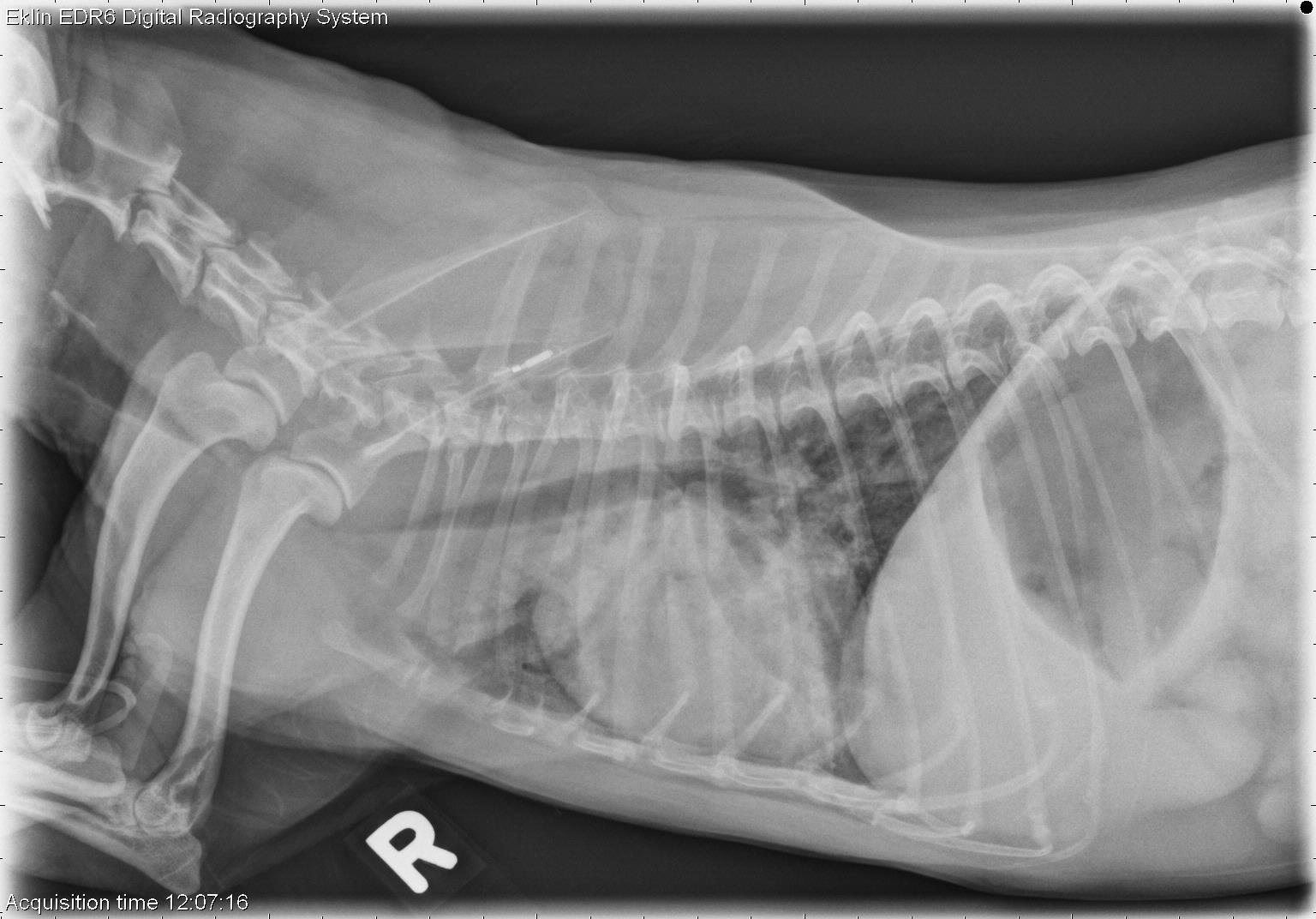
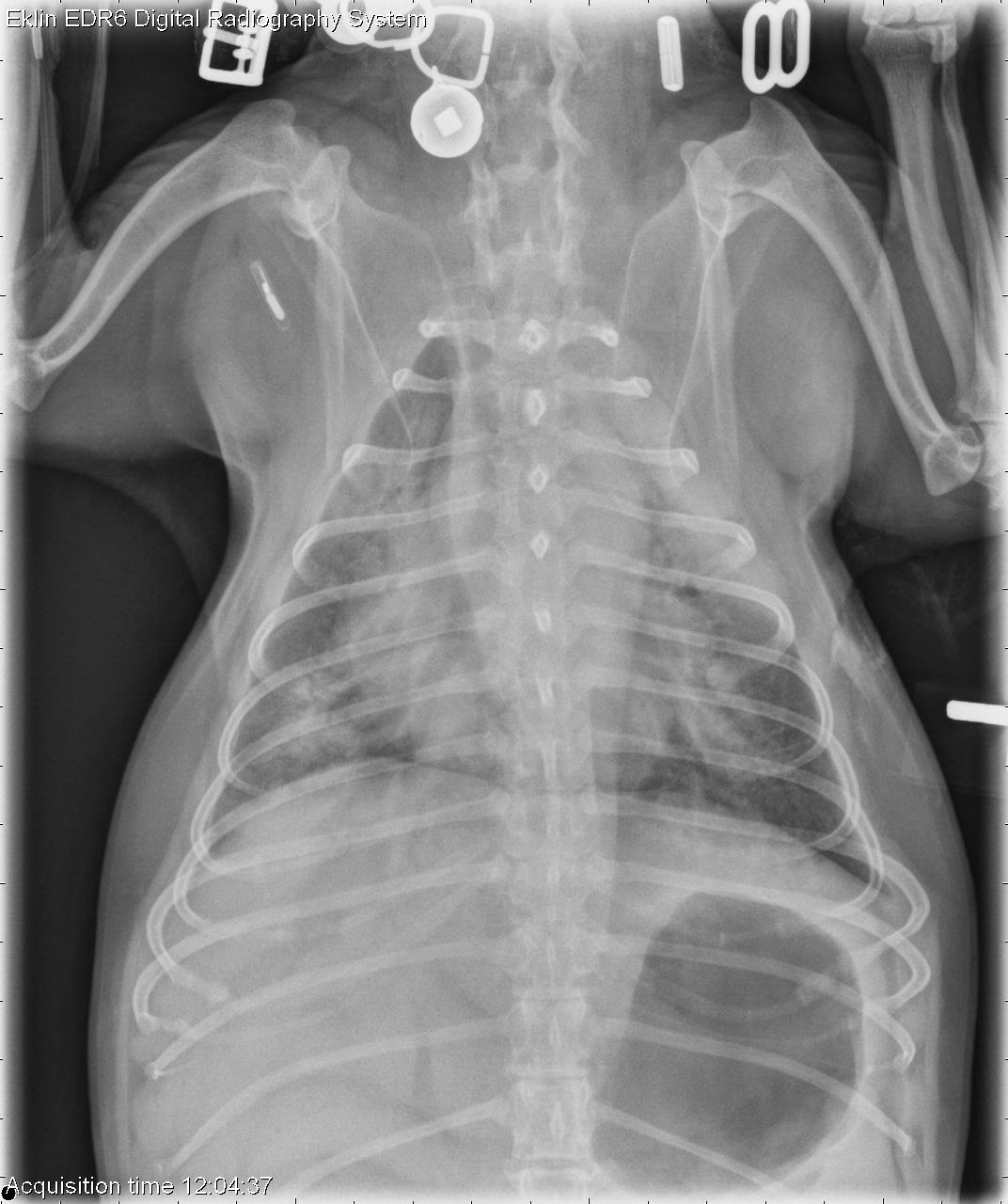
This week’s case is a 9-year-old female neutered Yorkshire Terrier with respiratory distress. What abnormalities do you see on the radiographs?
Findings
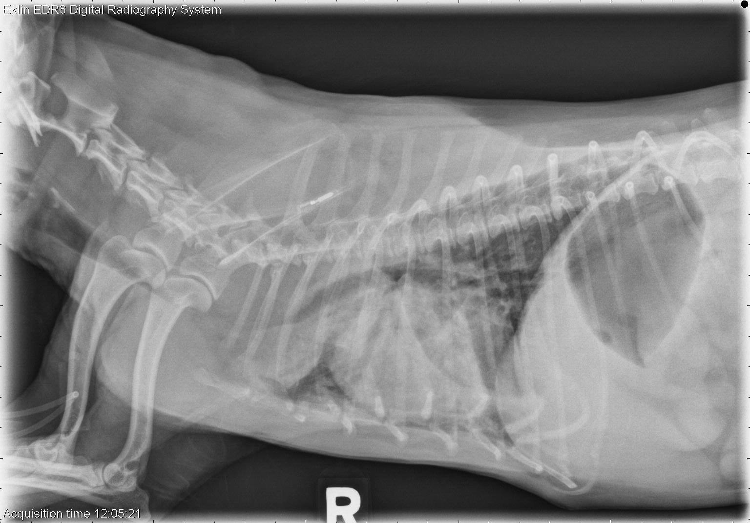
There is complete collapse of the trachea at the thoracic inlet with dynamic change in diameter in the thoracic portion. There is patchy alveolar infiltrate in all lung lobes, most apparent in the left cranial and right caudal lobes. The bronchi to the left cranial lobe are dilated and do not taper appropriately. The cardiac silhouette is moderately enlarged. There is gas in the stomach, likely secondary to aerophagia. The liver is enlarged and roundedDiagnosis
- Tracheal collapse (tracheomalacia), with a probable bronchial component and/or bronchiectasis
- Secondary pneumonia and/or non-cardiogenic pulmonary edema
- Hepatomegaly may be due to congestion, metabolic, inflammatory, toxic, or infiltrative disease