This week’s case is a 2-year-old male neutered French bulldog with central nervous system signs. These radiographs were taken prior to anesthesia.
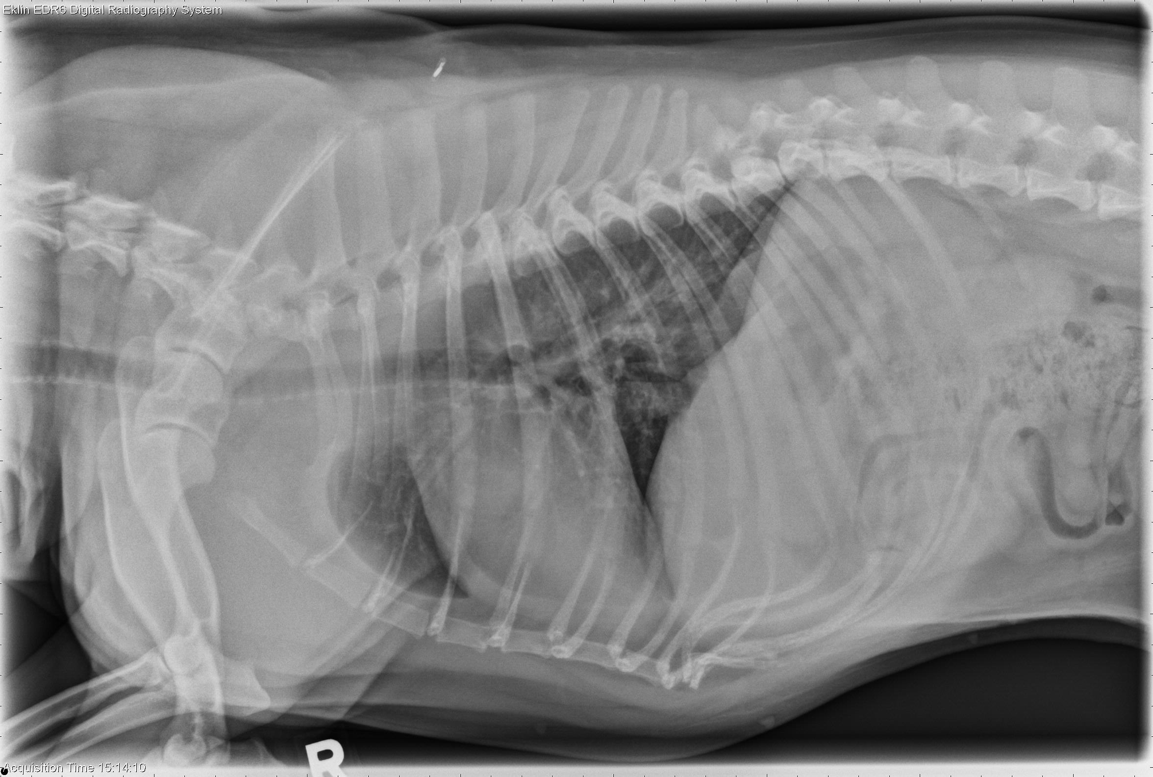
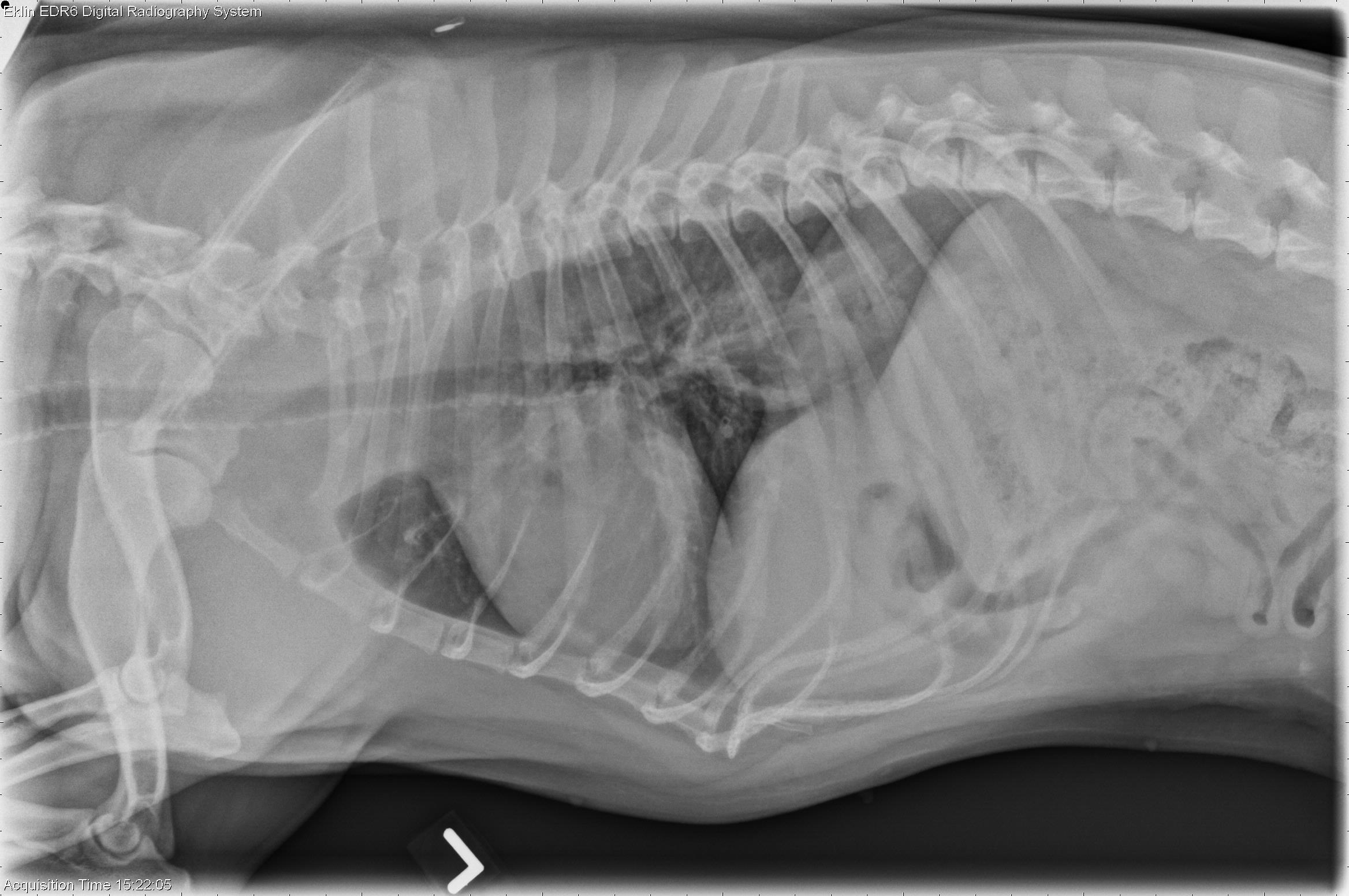
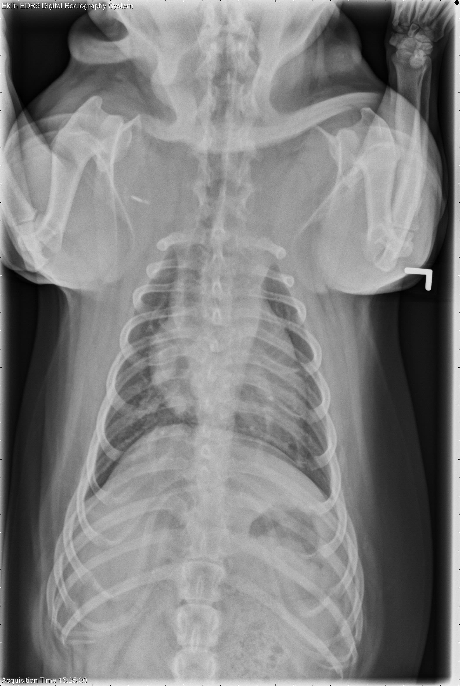
Findings
The cardiovascular structures are normal in size and shape. The airways are patent. There is a broad-based soft tissue and gas opacity at the level of the diaphragm on the left lateral projection. The pleura and mediastinum are normal. Hemivertebrae are present in the thoracic spine, consistent with breed.Differential Diagnosis
The intermittent soft tissue opacity in the region of the esophagus is most consistent with a sliding hiatal hernia, seen commonly in brachycephalic breeds.Diagnosis
- Sliding hiatal hernia (incidental, unlikely related to clinical signs)
- Inflammatory brain disease diagnosed on MRI