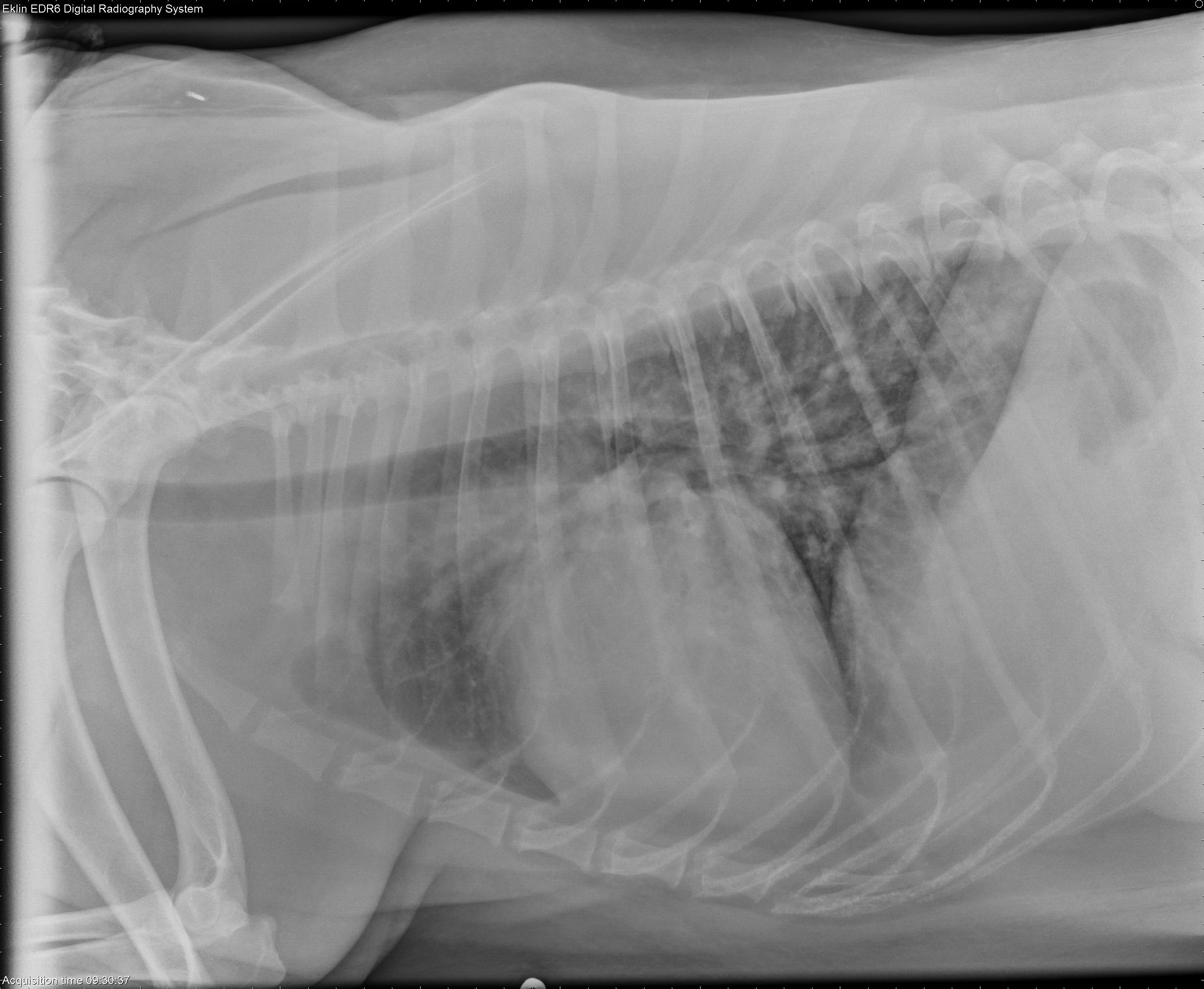
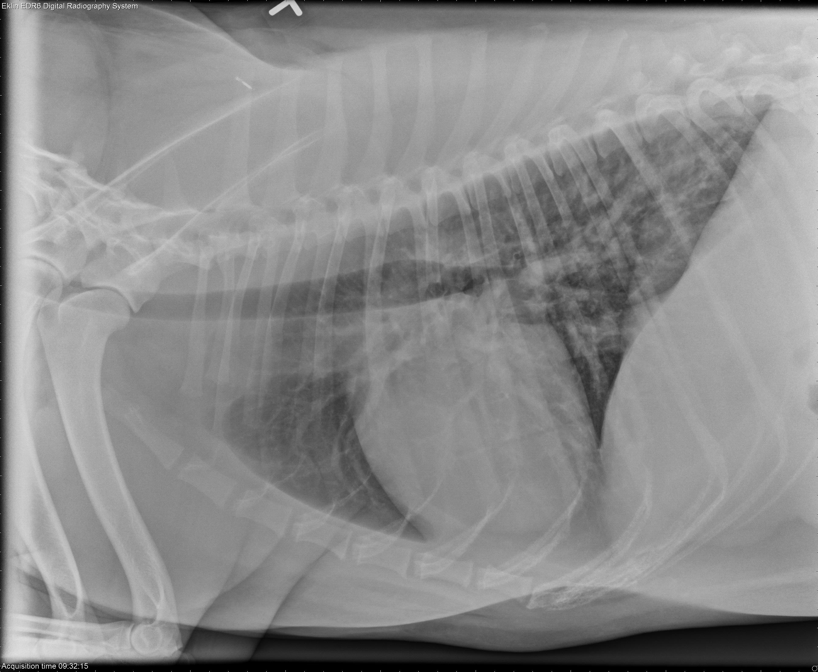
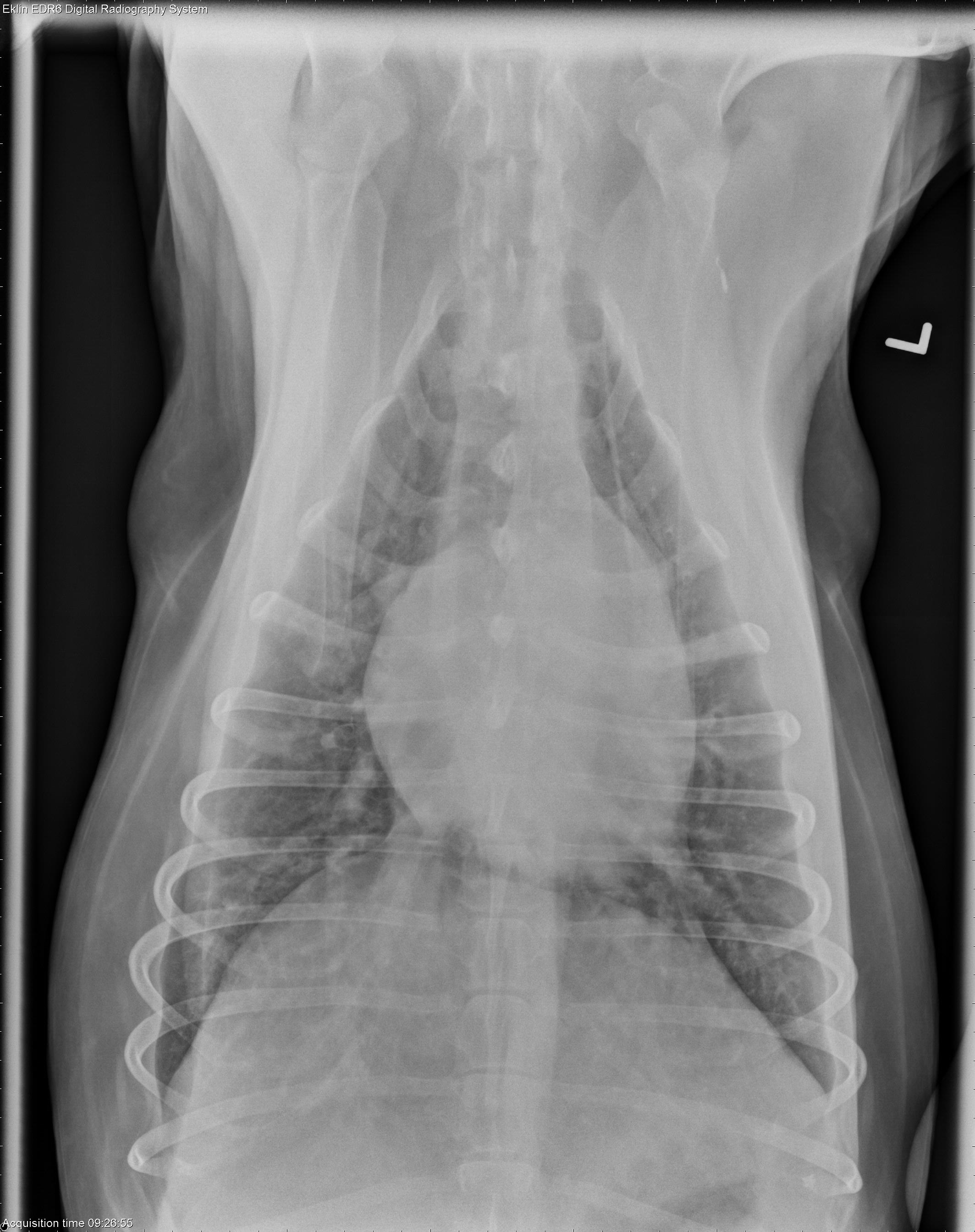
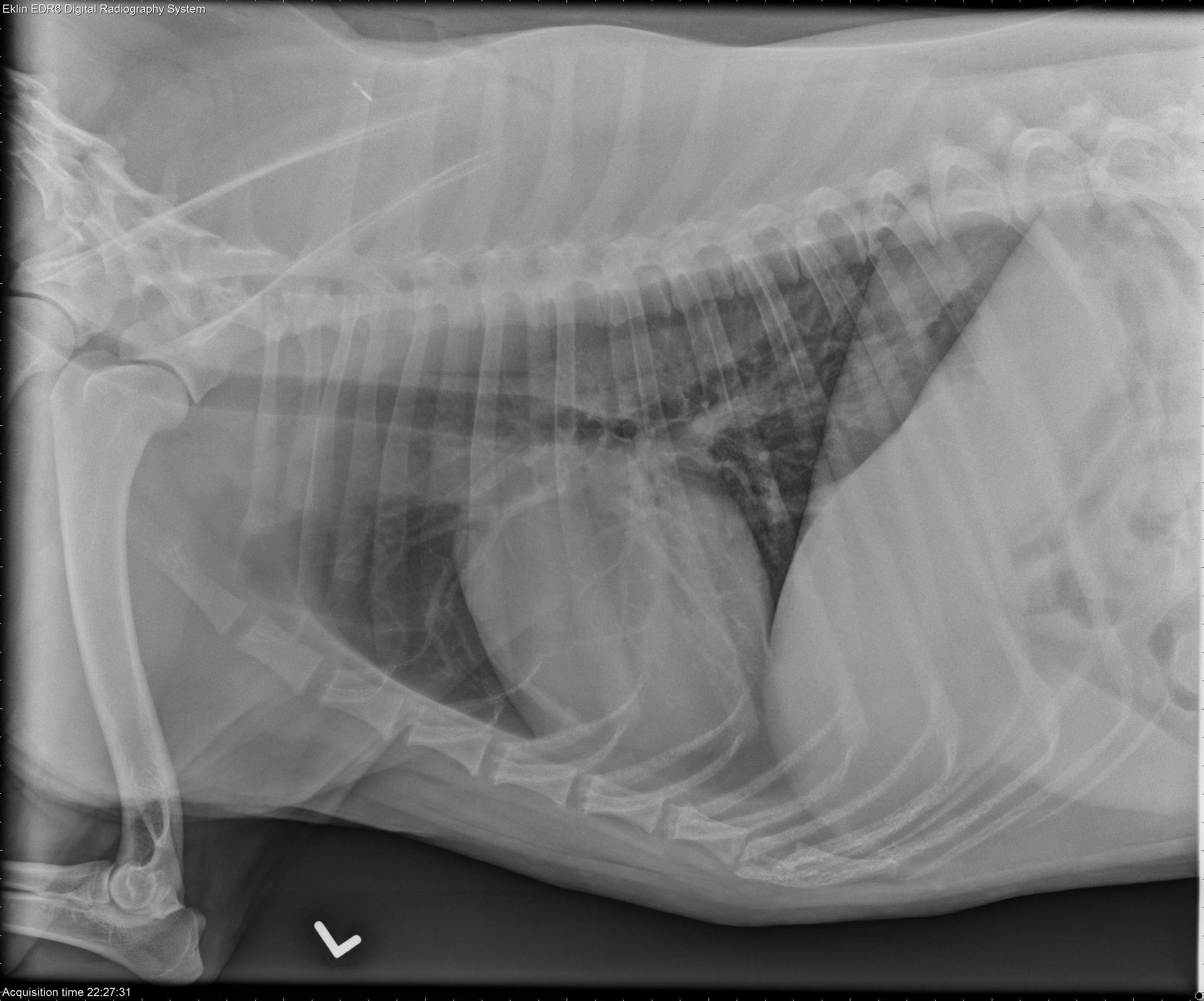
This week’s case is a 6-year-old female neutered Labrador Retriever with swelling after an allergy injection. Compare these radiographs to the second set of three images taken a few days prior.
Findings
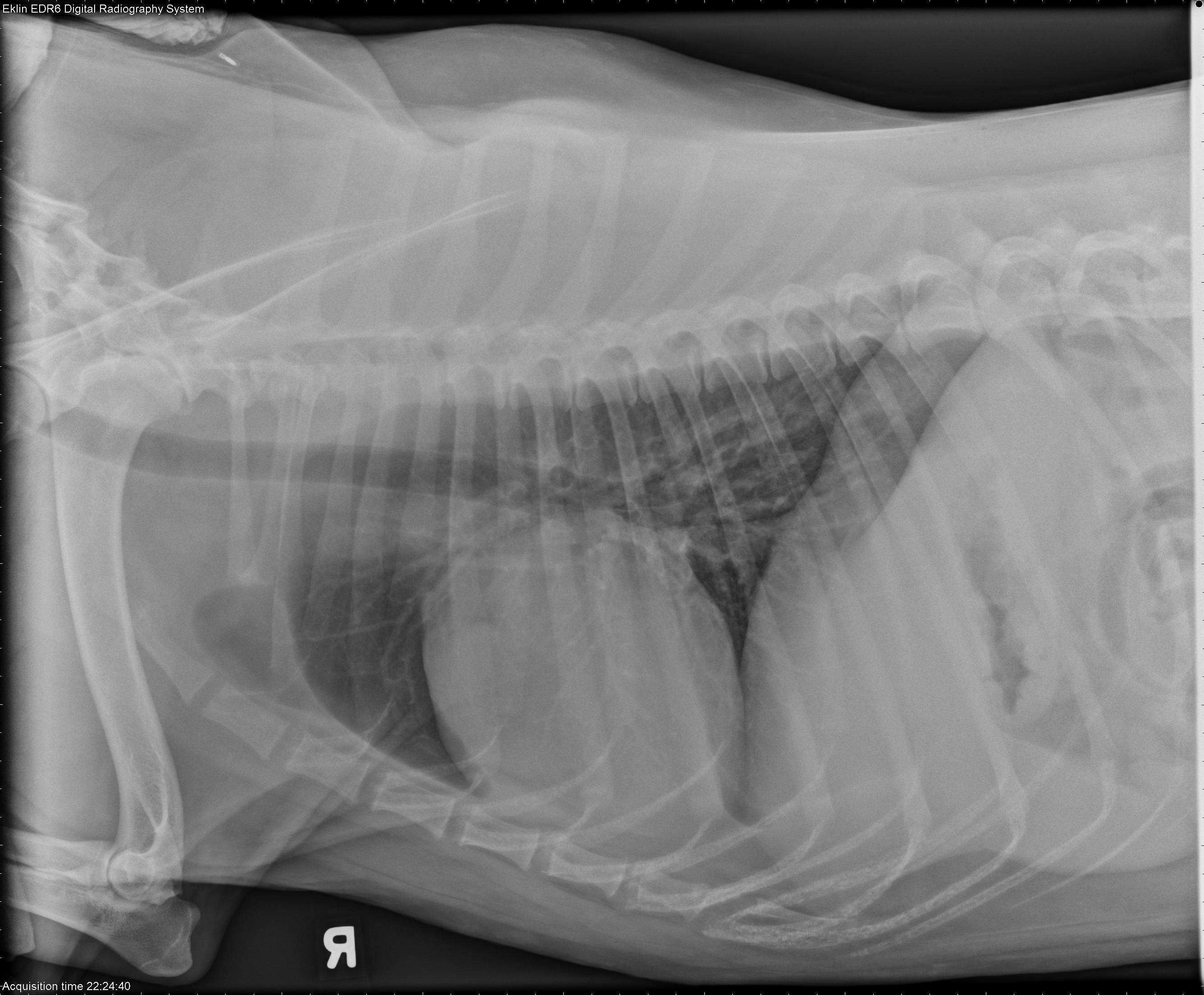
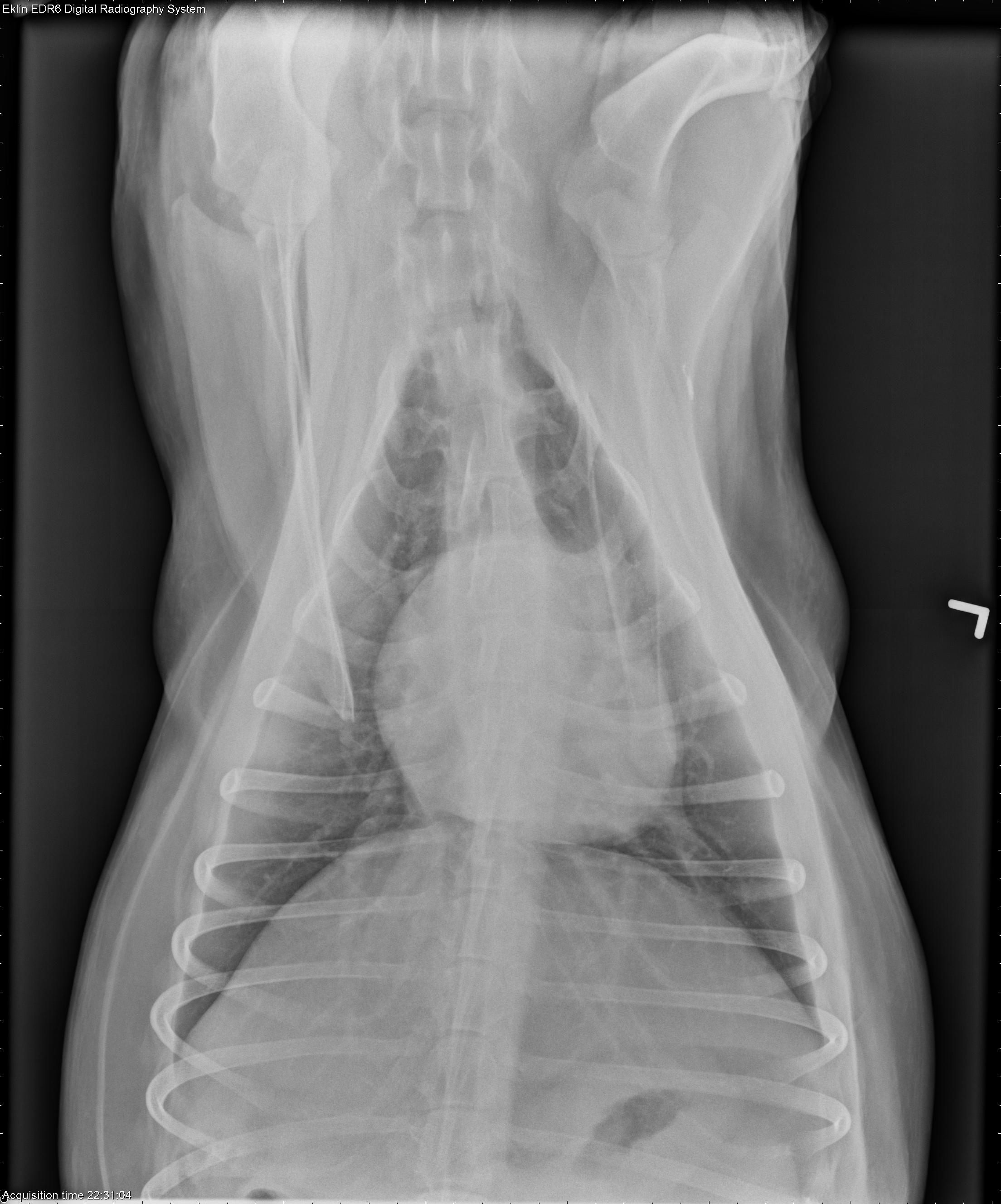
There is a diffuse bronchointerstitial pattern most prominent in the caudal lung lobes. Compared to several days previously (second set of images), this appear more pronounced. The heart is normal in size, however the pulmonary vasculature is enlarged. There are thin pleural fissure lines visible. The soft tissues of the ventral thoracic wall appear thickened.Differential Diagnosis
Non-cardiogenic edema and/or DIC secondary to immune mediated disease with acute exacerbation.Diagnosis
- Probable non-cardiogenic pulmonary edema and pleural effusion.
- Necropsy findings included a serosanguinous pleural effusion, but no evidence of microthrombi in the vasculature.