These radiographs are of a 9-year-old cat with labored breathing and panting. What are your differential diagnoses?
Findings
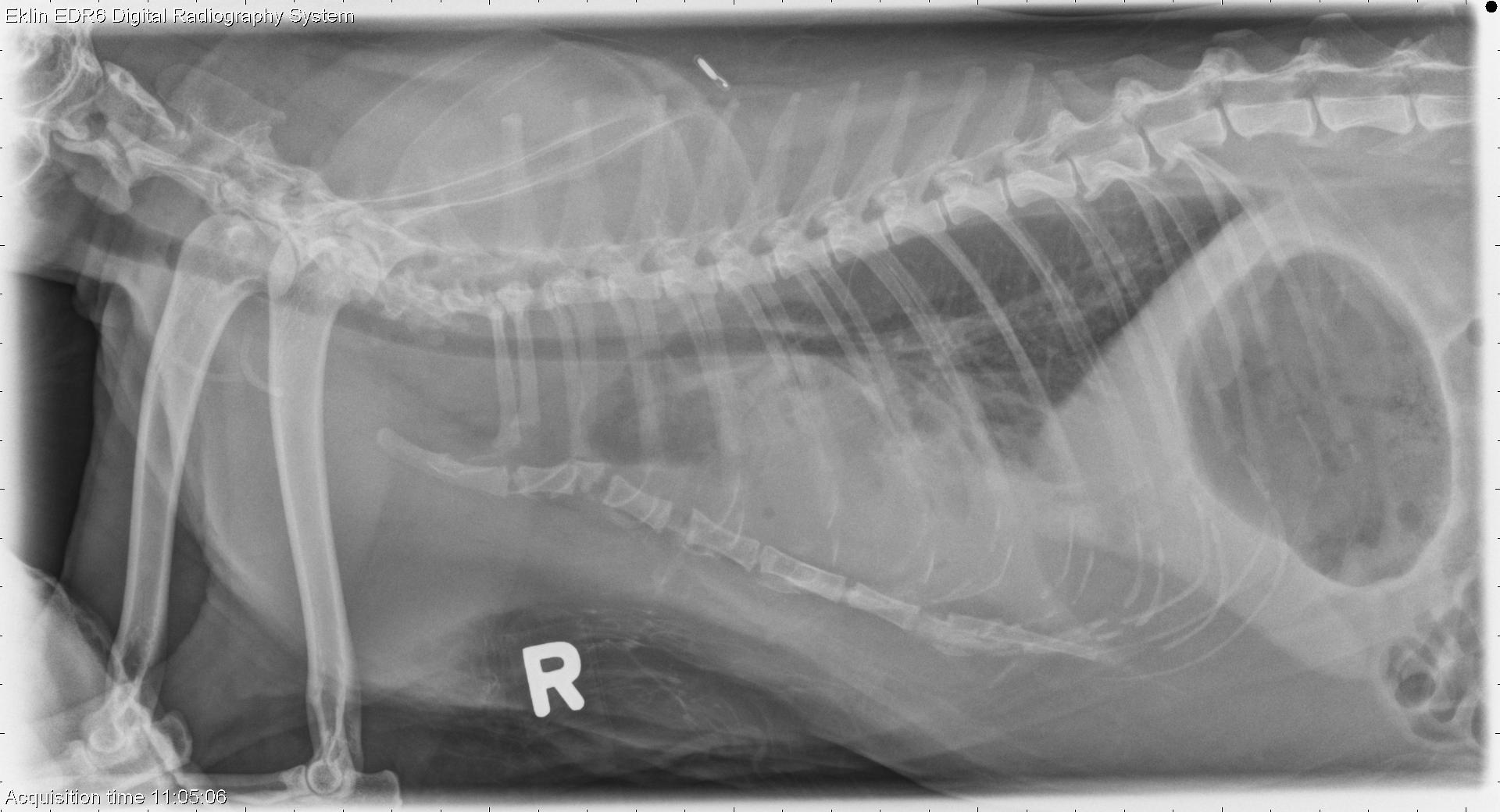
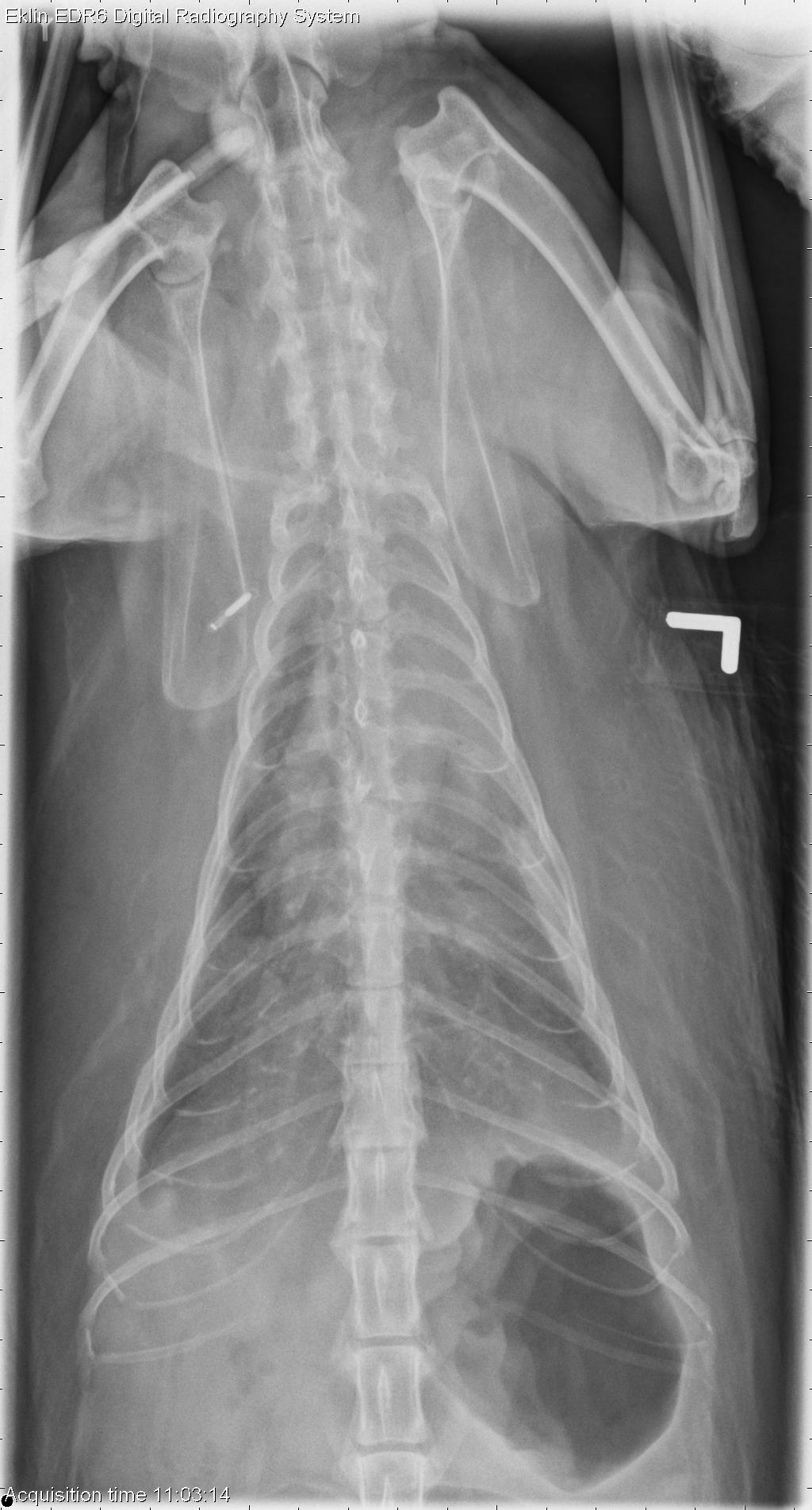
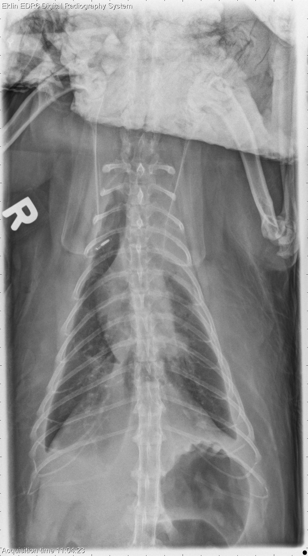
There is increased soft tissue opacity within the thorax, with retraction of the lung lobes and evidence of fissure lines. A few small gas bubbles are visible in the pleural space, secondary to thoracocentesis. The cardiac silhouette is obscured on the dorsoventral projection, but appears normal in size on the ventrodorsal projection. There is increased opacity and decreased volume of the left cranial lung lobe. A pulmonary nodule is also evident overlying the left side of the cardiac silhouette. Ill-defined nodules are present in the right caudal lung lobe.Differential Diagnosis
- Pulmonary mass (primary pulmonary neoplasia, granuloma)
- Possible metastatic disease or diffuse granulomatous disease
- Secondary pleural effusion and atelectasis
Diagnosis
- Adenocarcinoma with intrapulmonary metastasis
- Chronic proliferative pleuritis and pleural fibrosis and atelectasis, secondary to pleural effusion