This week’s case is a 12-year-old male neutered Domestic Shorthair Cat with 2-week history of increased respiratory effort and several days of inappetence. The feline thorax can be difficult to interpret. Can you find the abnormality and list some differential diagnoses?
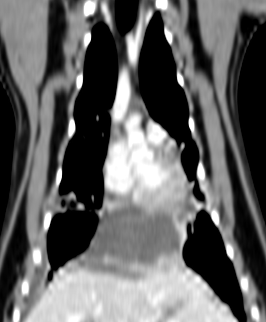
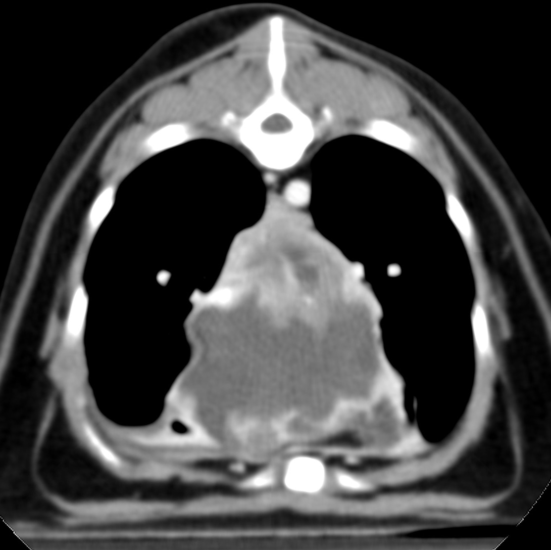
Findings
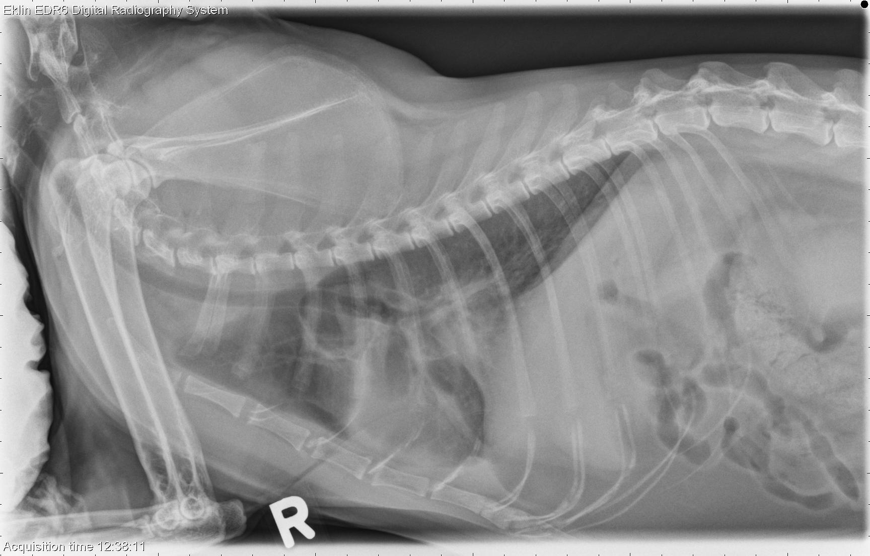
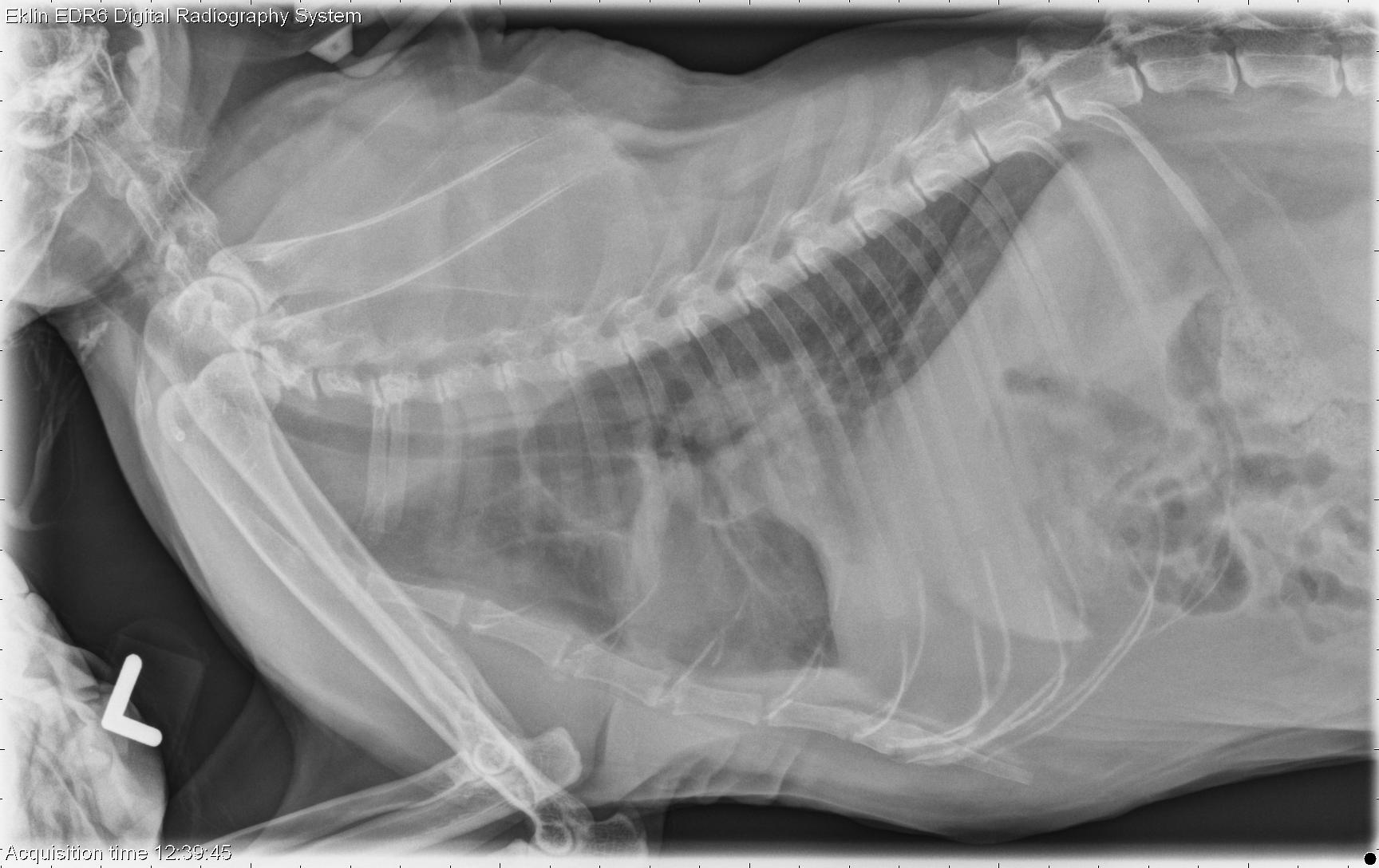
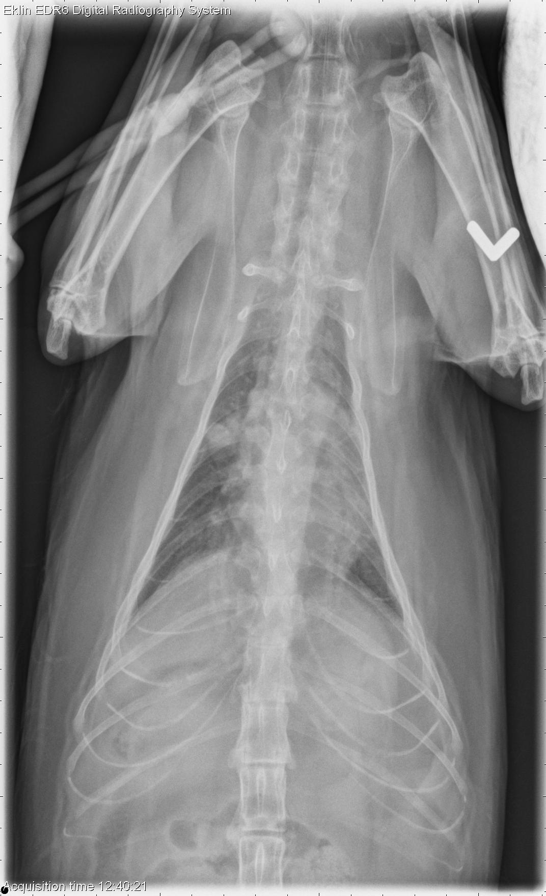
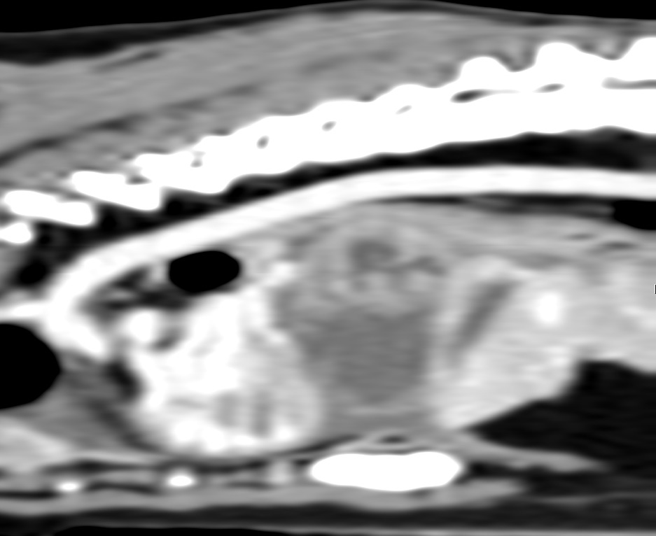
The cardiac silhouette is normal in size and shape, and the pulmonary vasculature is normal. There is an increased soft tissue opacity in the region of the accessory lung lobe or caudal mediastinum, that silhouettes with the diaphragm and obscures the caudal cardiac silhouette. Soft tissue opacity fissure lines are visible between several lung lobes. There is a mild bronchial pattern throughout the lungs. No significant abnormalities are seen in the musculoskeletal structures.Differential Diagnosis
- Mass in the accessory lung lobe or caudal mediastinum (neoplasia, granuloma, abscess)
- Pleural effusion (transudate, exudate, blood, chyle), likely secondary to the mass
- Bronchial pattern may be inflammatory or represent mild edema