This week’s case is a 16-year-old Australian Cattle Dog with episodes of collapse. What are your differential diagnoses?
Findings
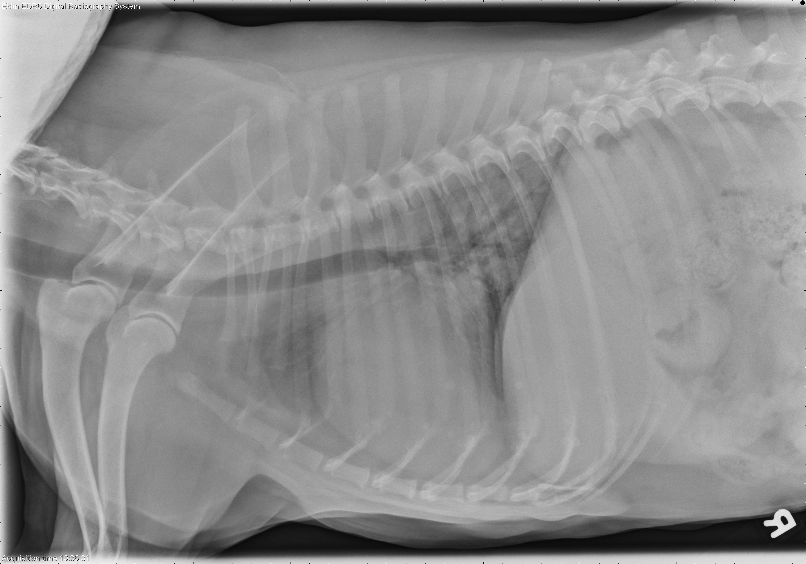
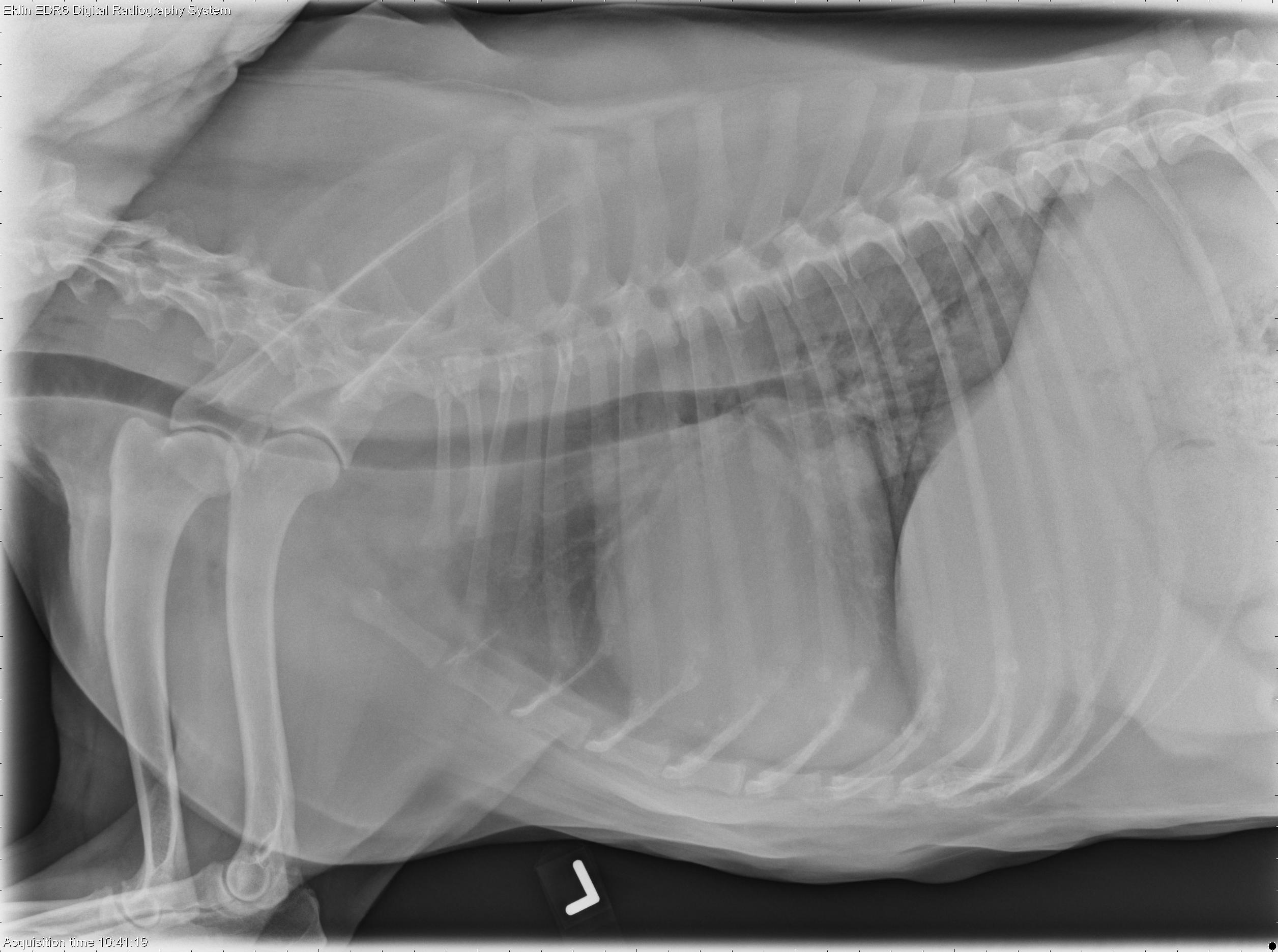
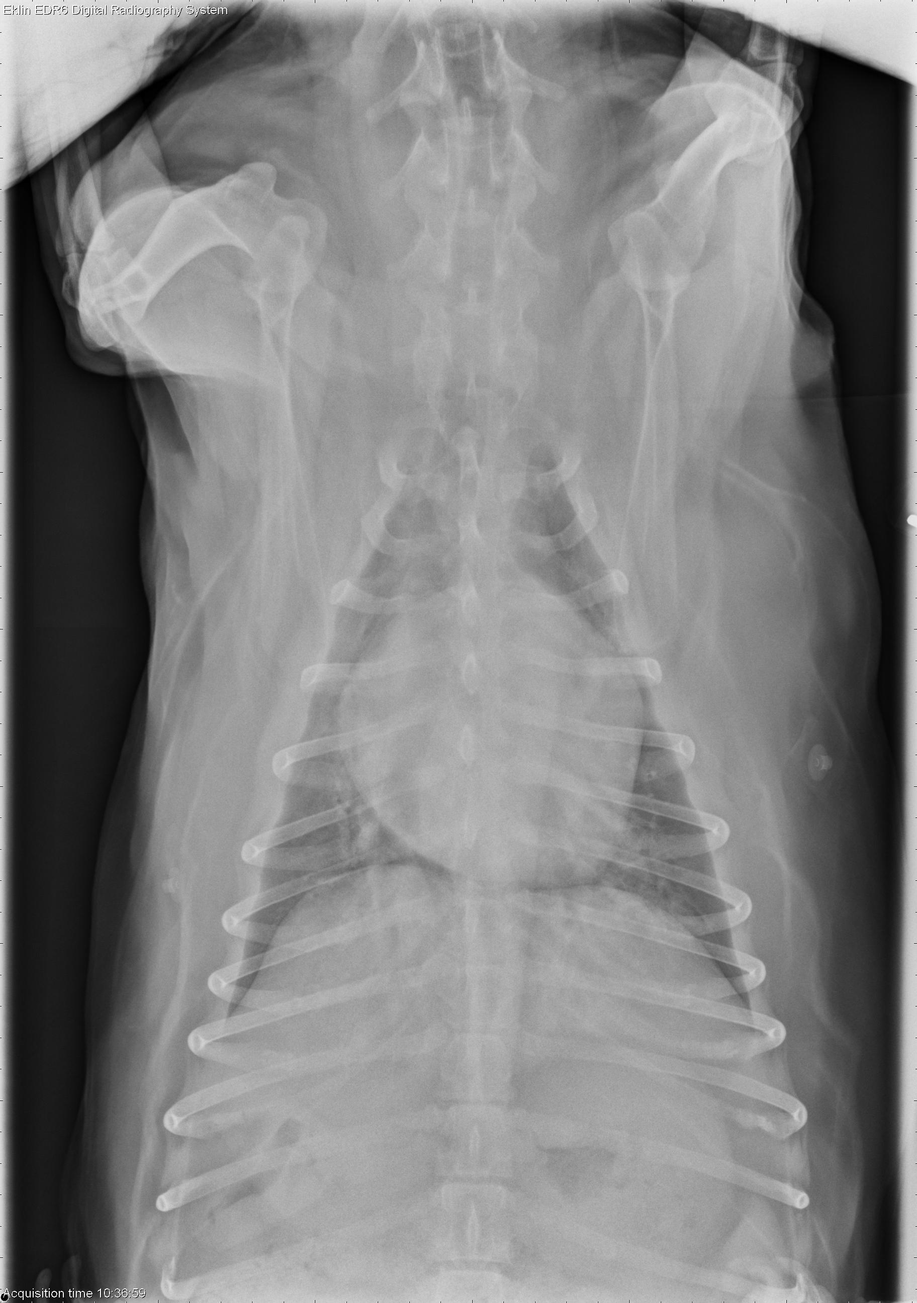
The cardiac silhouette is enlarged, with marked left atrial enlargement. The trachea is elevated parallel to the spine, and there is a straightening of the caudal cardiac margin. The pulmonary vasculature is normal in size. There is a fine interstitial pulmonary pattern throughout the lungs, most concentrated in the caudal lung lobes. No pleural effusion is evident. ECG pads are visible on the left and right body wall, causing round opacities superimposed over the heart on the lateral projections. In the portion of the abdomen included, the liver extends beyond the costal arch. The caudal vena cava is normal in size, and the peritoneal detail is normal.Differential Diagnosis
- Cardiomegaly and left heart failure, likely secondary to valvular degeneration
- Hepatomegaly may be due to congestion, metabolic, inflammatory, toxic, or infiltrative disease.
Diagnosis
- Severe decompensated myxomatous mitral valve degeneration and mitral valve chordae tendineae rupture
- Pulmonary hypertension and pulmonary fibrosis secondary to left heart failure