This week’s case is a 12-year-old dog that presents for acute onset of vomiting. There are many findings in this case. Can you see all of them?
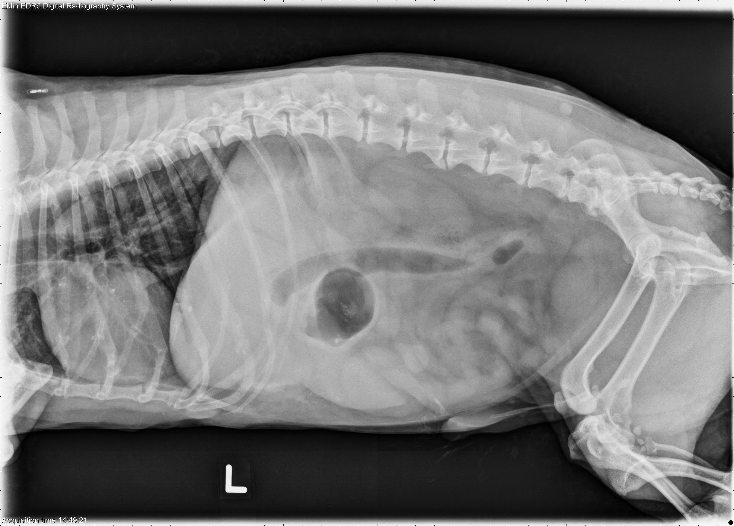
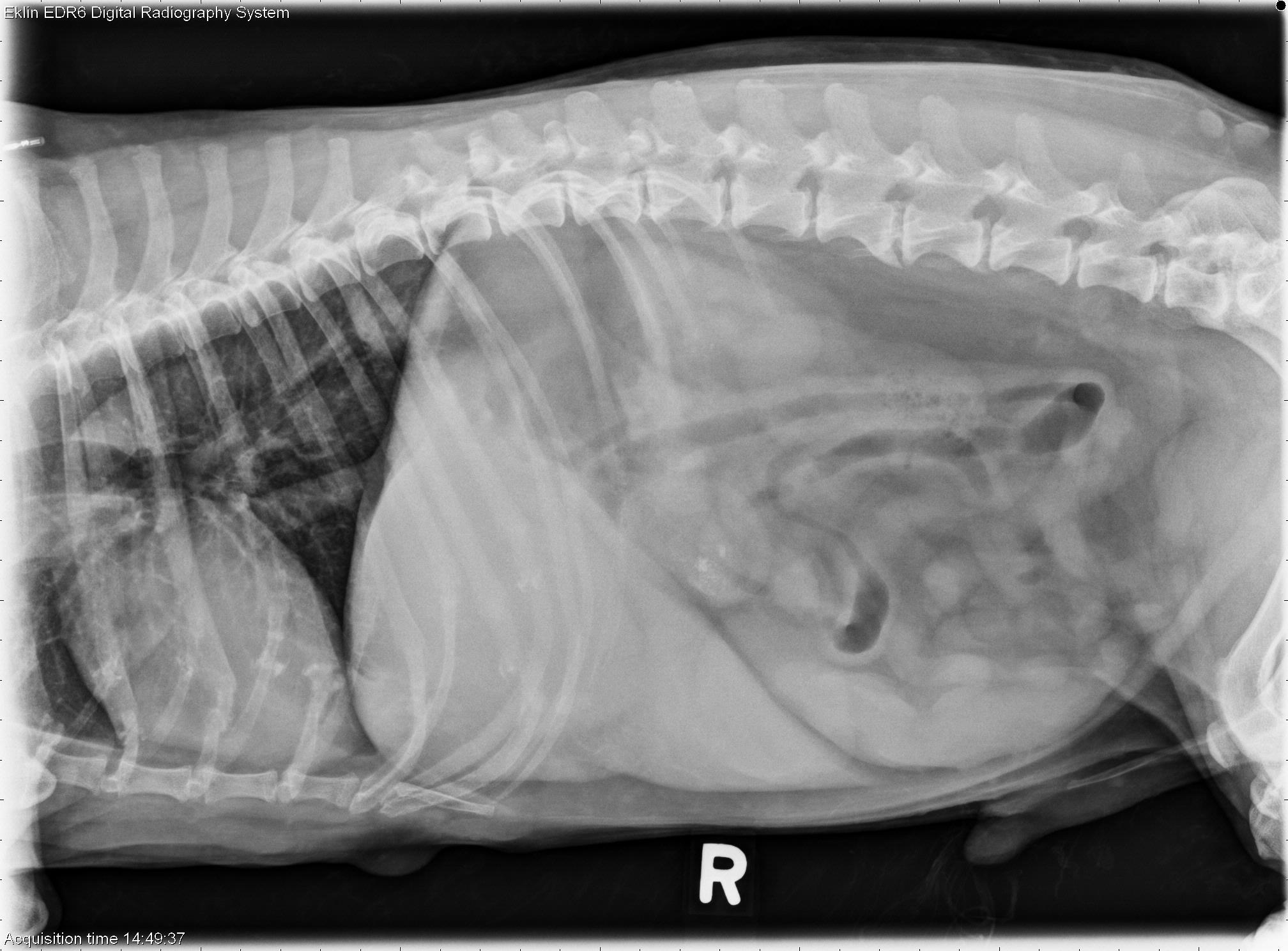
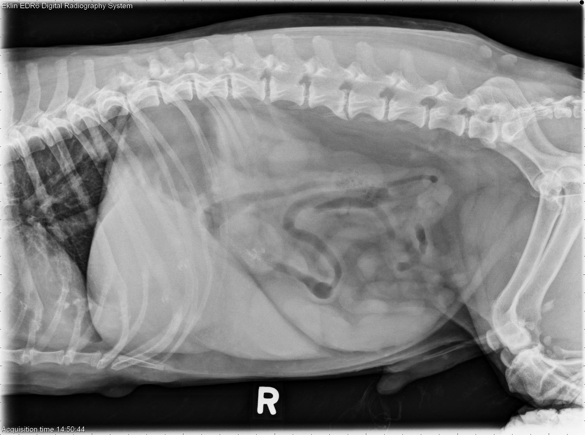
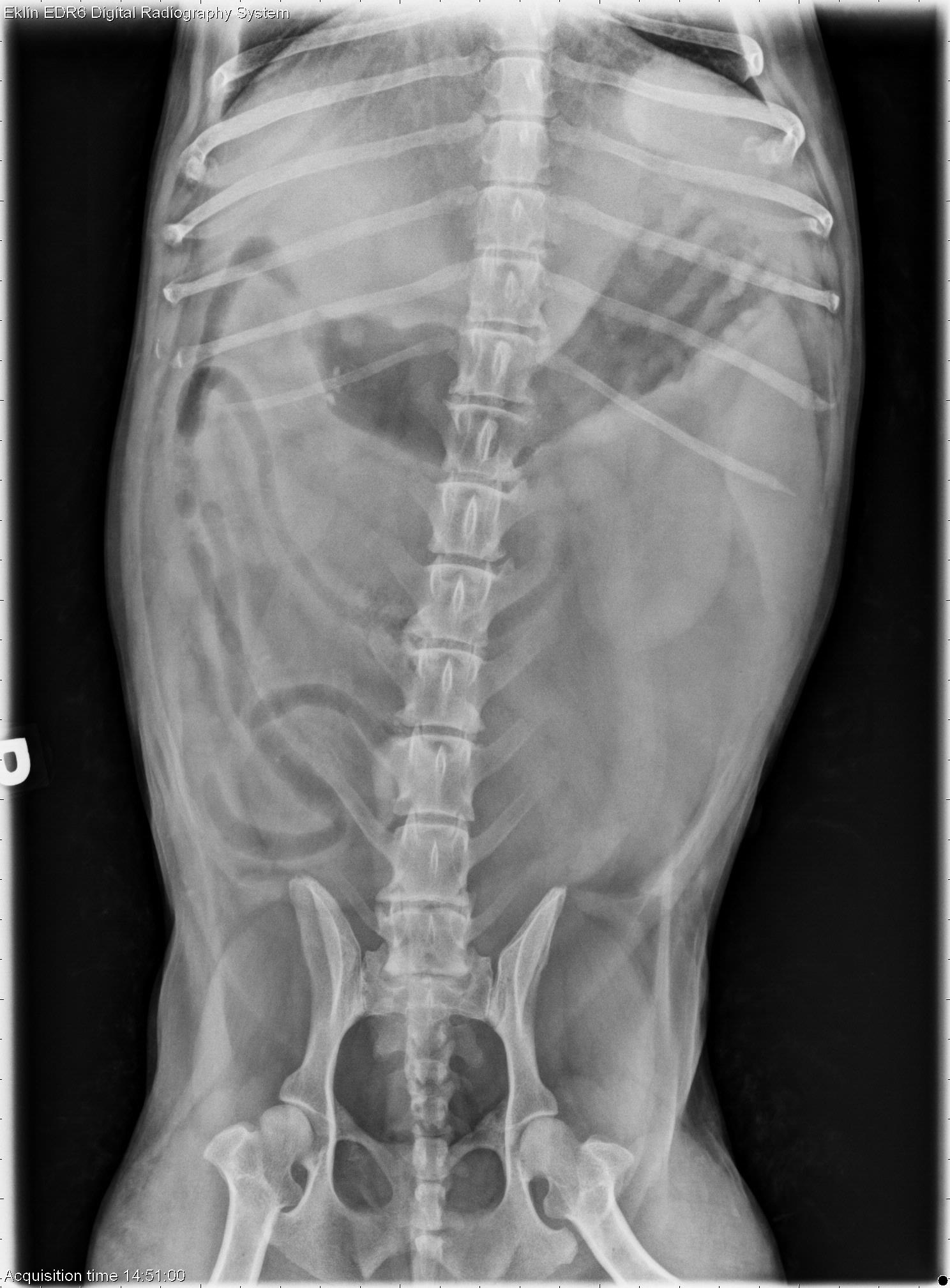
Findings
Abdomen: There is mild abdominal distention and poor detail in the cranioventral abdomen. The liver is enlarged and extends beyond the costal arch. There is a round soft tissue mass superimposed over the caudoventral liver and spleen on the lateral projections. There is widening of the gastroduodenal angle on the ventrodorsal projection, and the duodenum is distended with gas on all projections. The kidneys are normal in size and shape. The bladder is normal. There are small mineral opacities in the region of the urethra caudal to the os penis. The facets of T12-L1 are absent on the left side due to previous laminectomy. In the portion of the thorax included, there is a round soft tissue mass in the left caudal lung lobe.Differential Diagnosis
- Abdominal mass – DDx liver, gall bladder, spleen, lymph nodes.
- Hepatomegaly – DDX inflammatory, metabolic, neoplastic, degenerative.
- Pancreatitis with effusion
- Urethral calculi
- Pulmonary mass – DDX primary pulmonary neoplasia, granuloma.
Diagnosis
Ultrasound and cytology findings included:- Gall bladder mucocele with bile peritonitis
- Hepatic mass (not sampled)
- Severe pancreatitis with mesenteric inflammation