This week’s case is a 10-year-old female neutered mixed breed dog with an ocular mass. This dog had a metastasis check after discovery of a pulmonary mass. What are your findings?
Findings
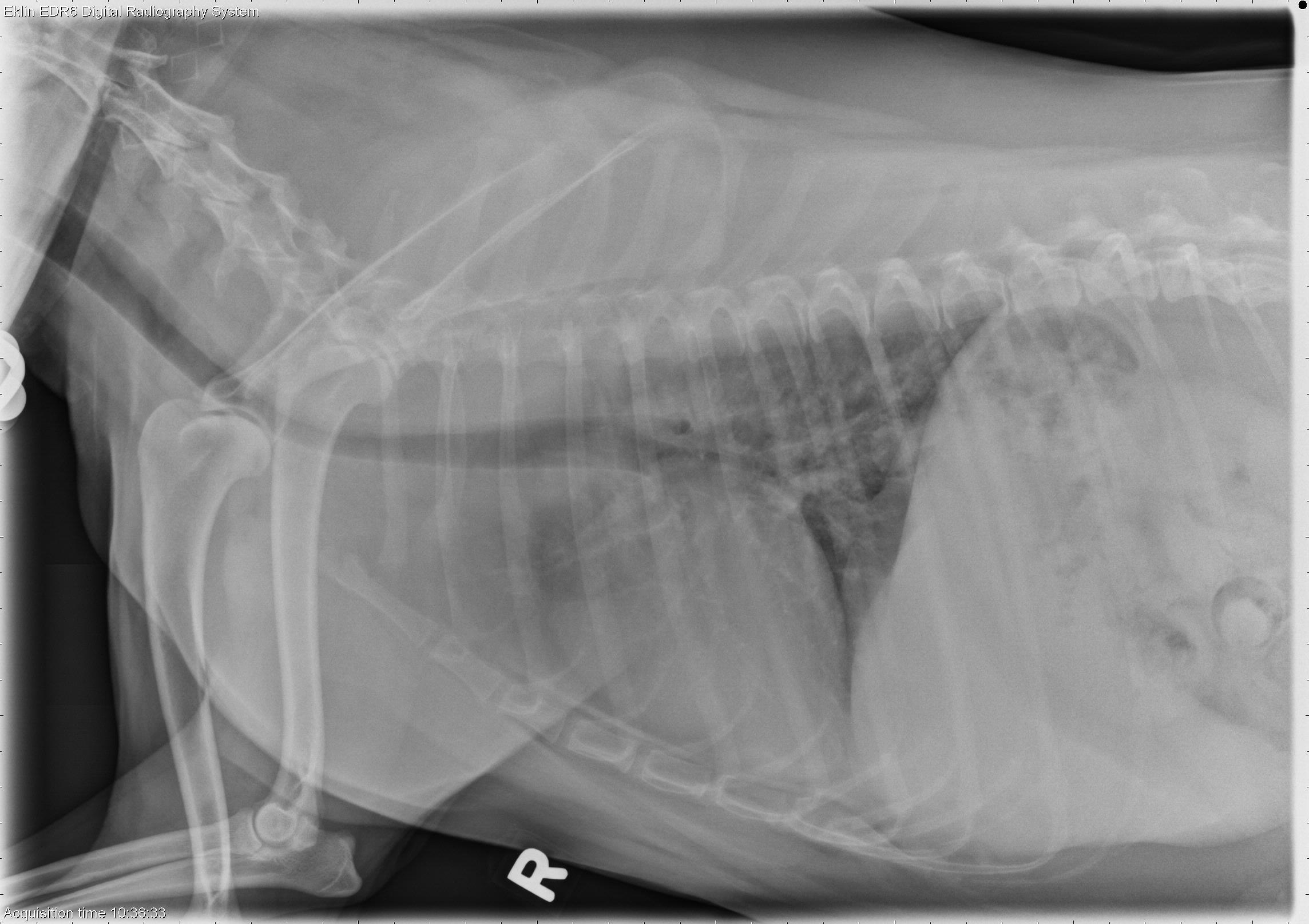
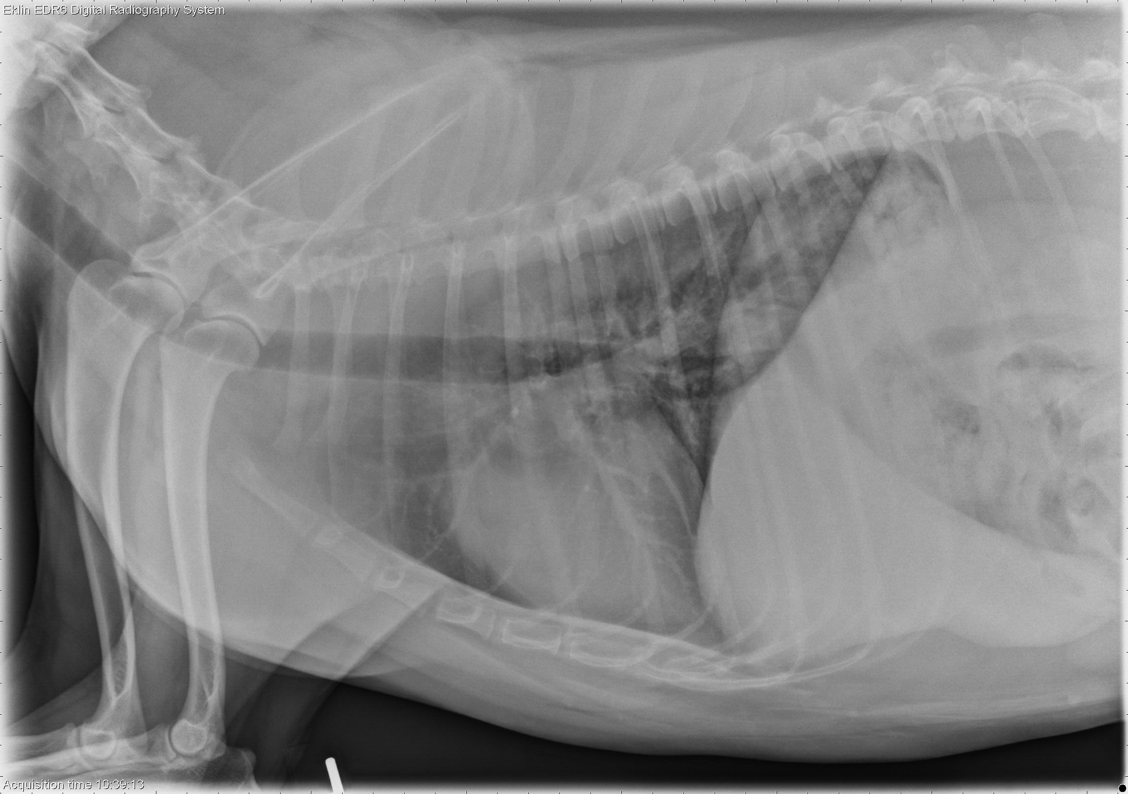
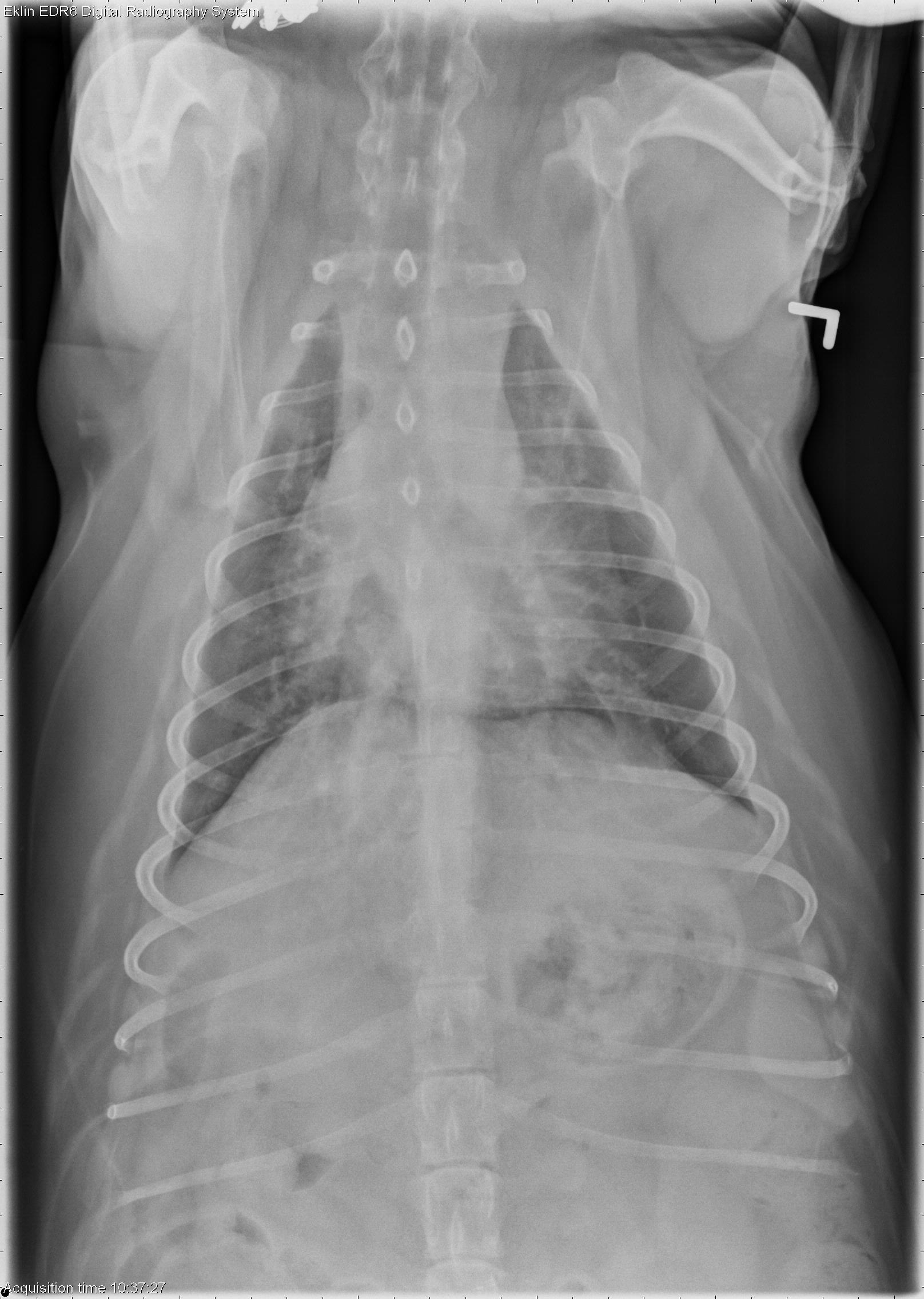
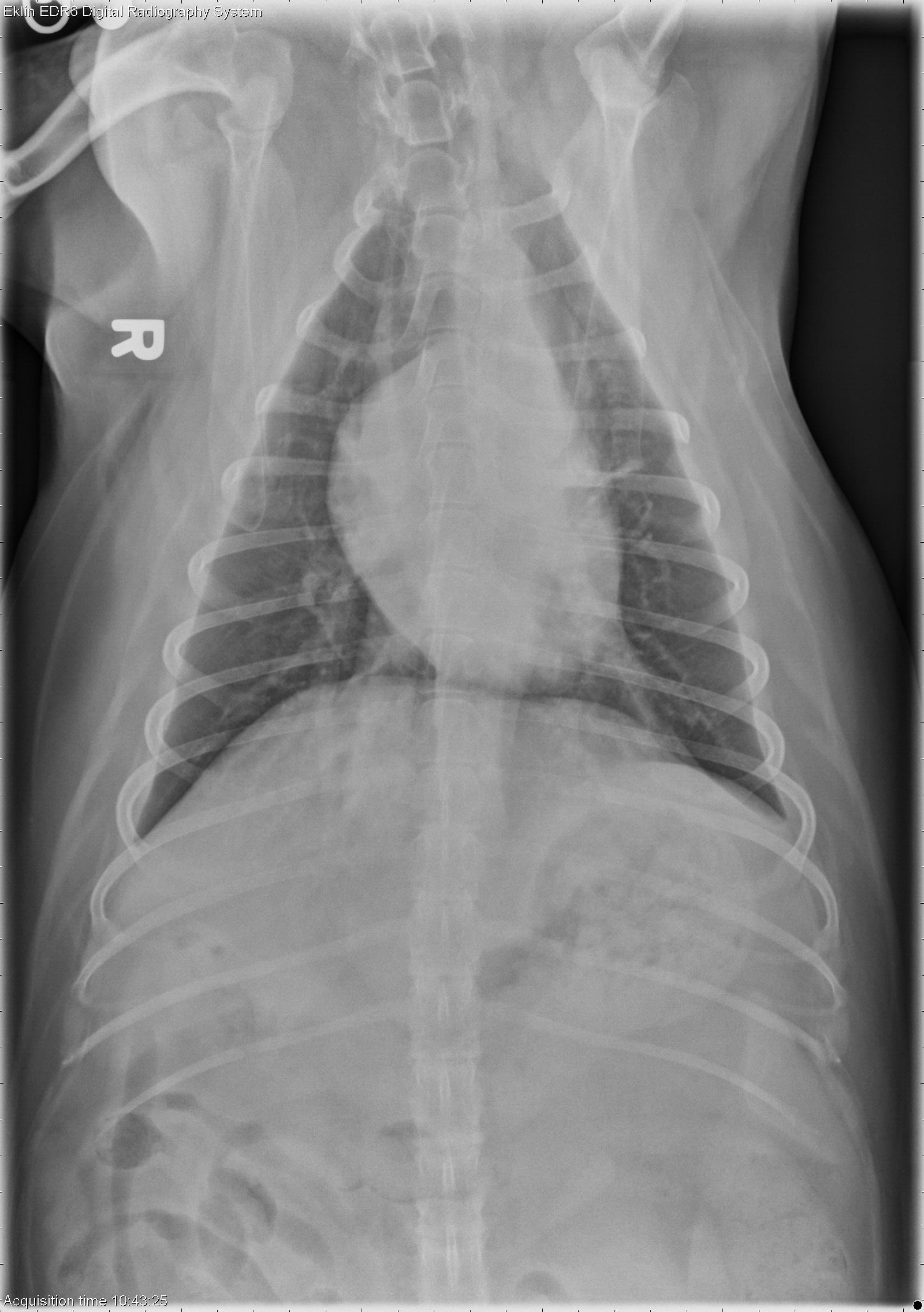
On the right lateral projection, there is a rounded soft tissue nodule dorsal to the caudal vena cava, superimposed on the crura of the diaphragm. The nodule is superimposed over the fluid-filled esophagus on the left lateral projection. The nodule is again seen on the ventrodorsal projection superimposed on the caudal vena cava to the right of the spine. The remainder of the pulmonary parenchyma is normal. No abnormalities are noted in the pleural space or mediastinum. The cardiovascular structures are normal. The liver is moderately enlarged.Differential Diagnosis
- Solitary pulmonary mass (metastasis vs. granulomatous)
- Hepatomegaly may be due to metabolic, inflammatory, toxic, or infiltrative disease.
Diagnosis
- Ocular nerve and intracranial meningioma
- Granuomatous pneumonia (Paragonimus sp. trematode)