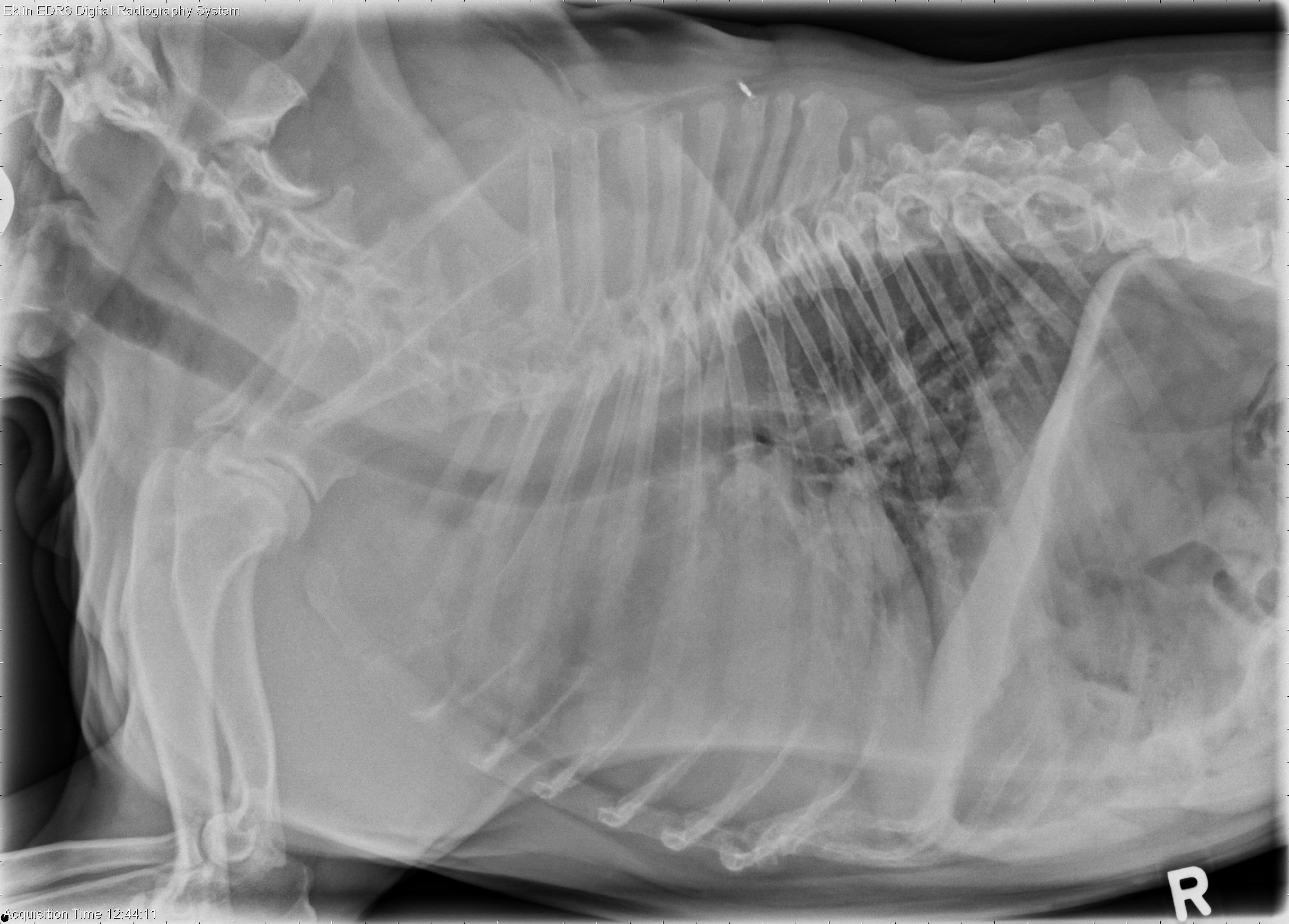
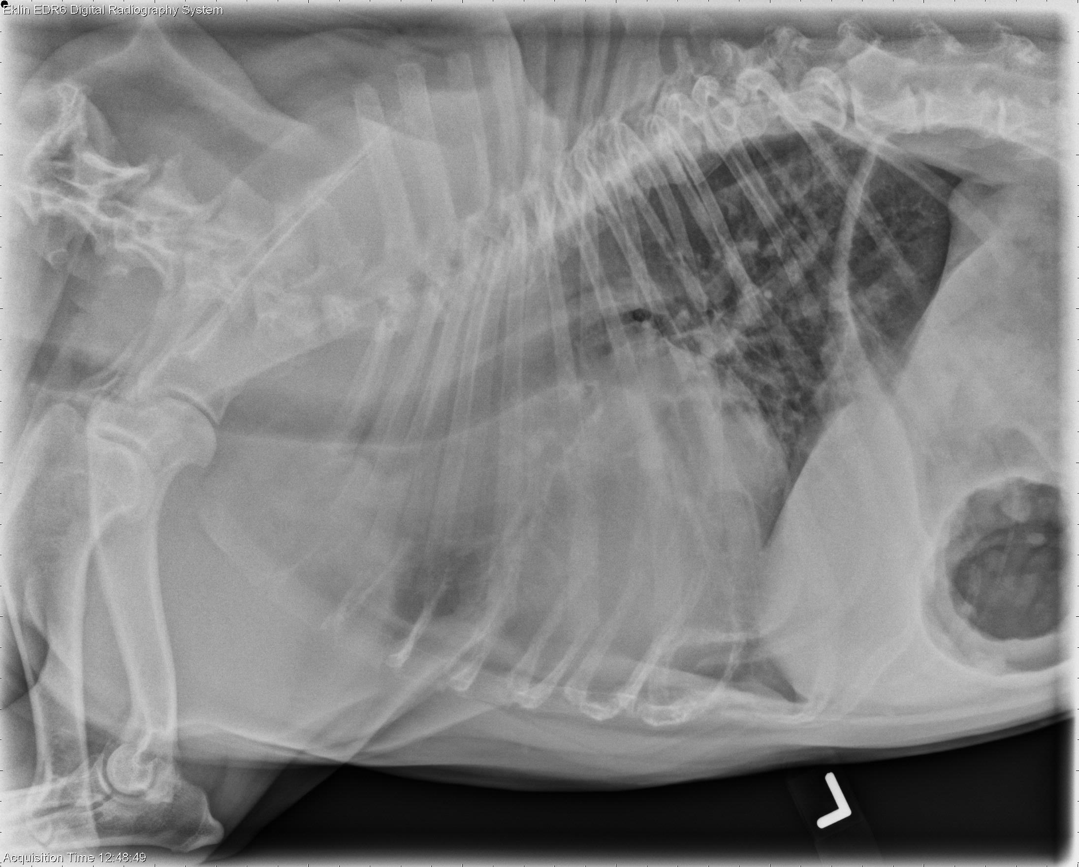
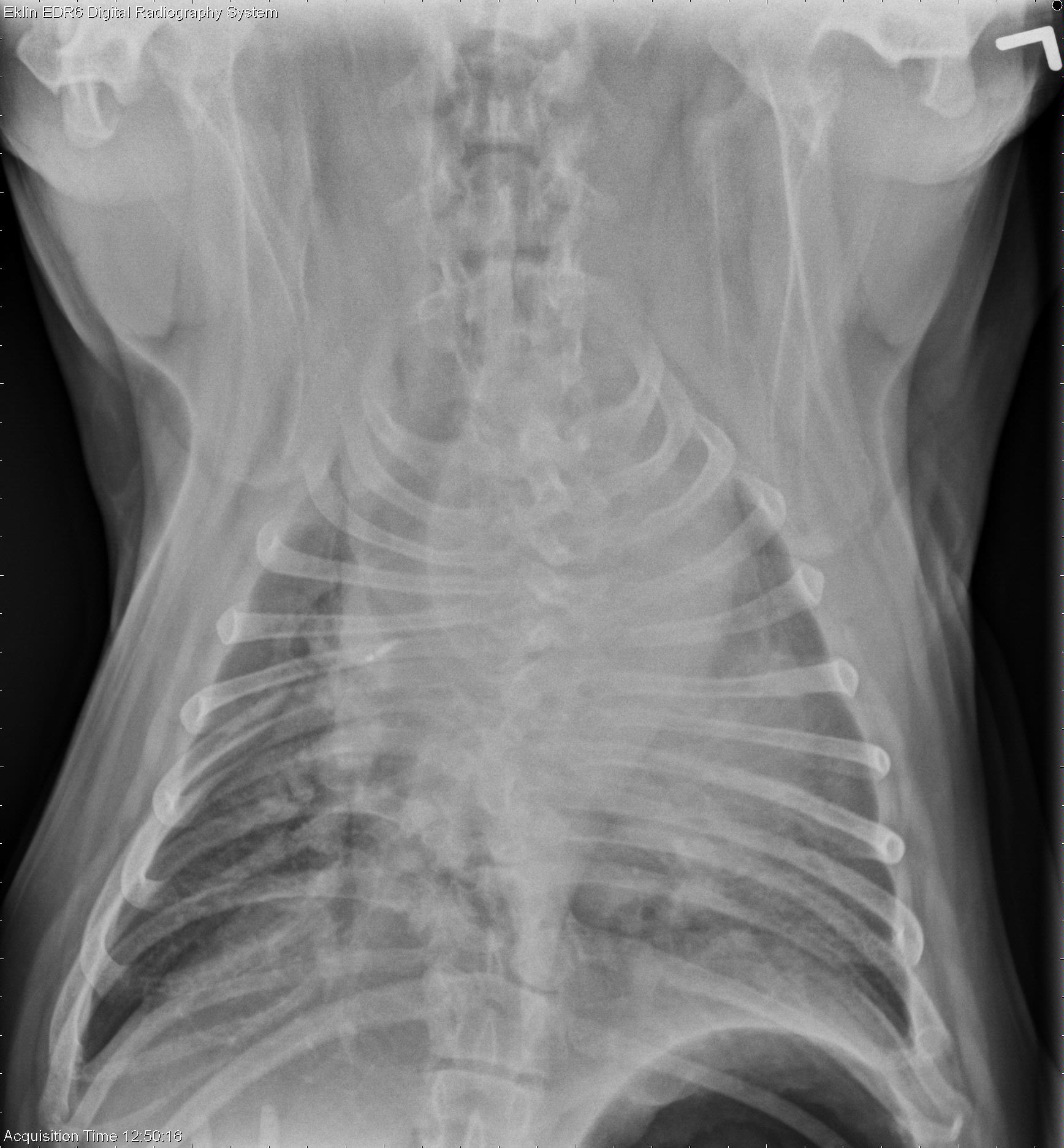
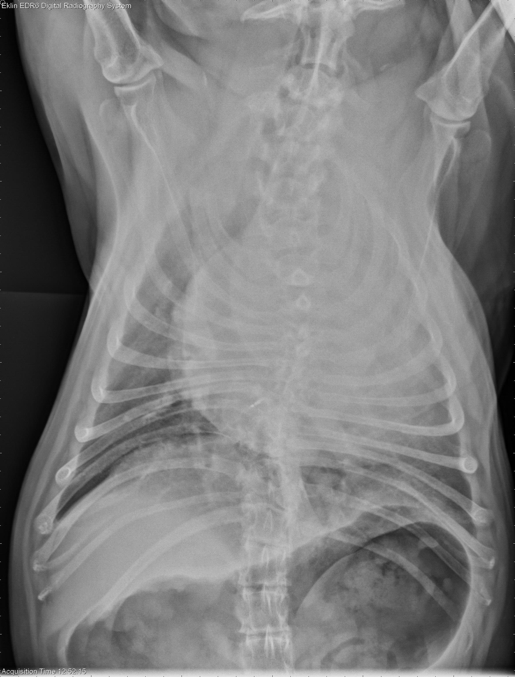
This week’s case is a 7-year-old female neutered Bulldog with periods of increased respiratory rate for 1 week. Thoracic radiographs of a bulldog are always challenging to interpret, but what are your findings?
Findings
On the lateral images, there is increased opacity in the cranial mediastinum. The trachea is normally positioned. There is a silhouette sign with the cranial cardiac silhouette. On the ventrodorsal projection, the cranial mediastinum bulges laterally and is markedly widened. The cardiac silhouette is mildly enlarged. There are congenital vertebral anomalies and spondylosis deformans in the thoracic spine. The liver appears small.Diagnosis
- Heart base mass at aortic root, unknown type
- Mild cardiomegaly with valvular degeneration and supraventricular tachycardia