Today’s case is an 18-month-old Rat Terrier who was kicked by a horse several months ago. Presents for respiratory distress. Challenge yourself to rank your differential diagnoses.
Findings
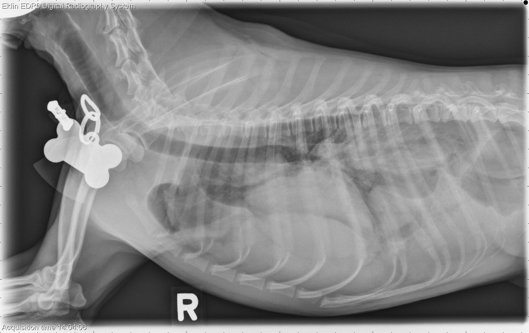
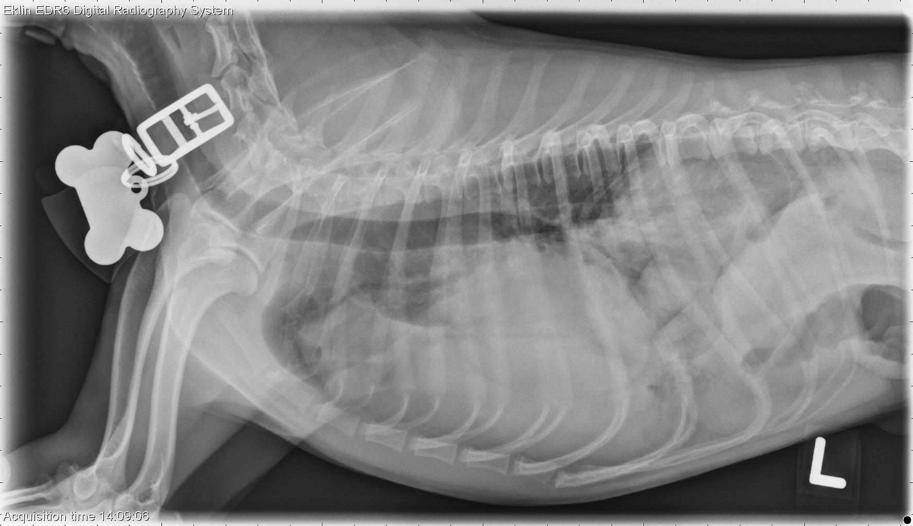
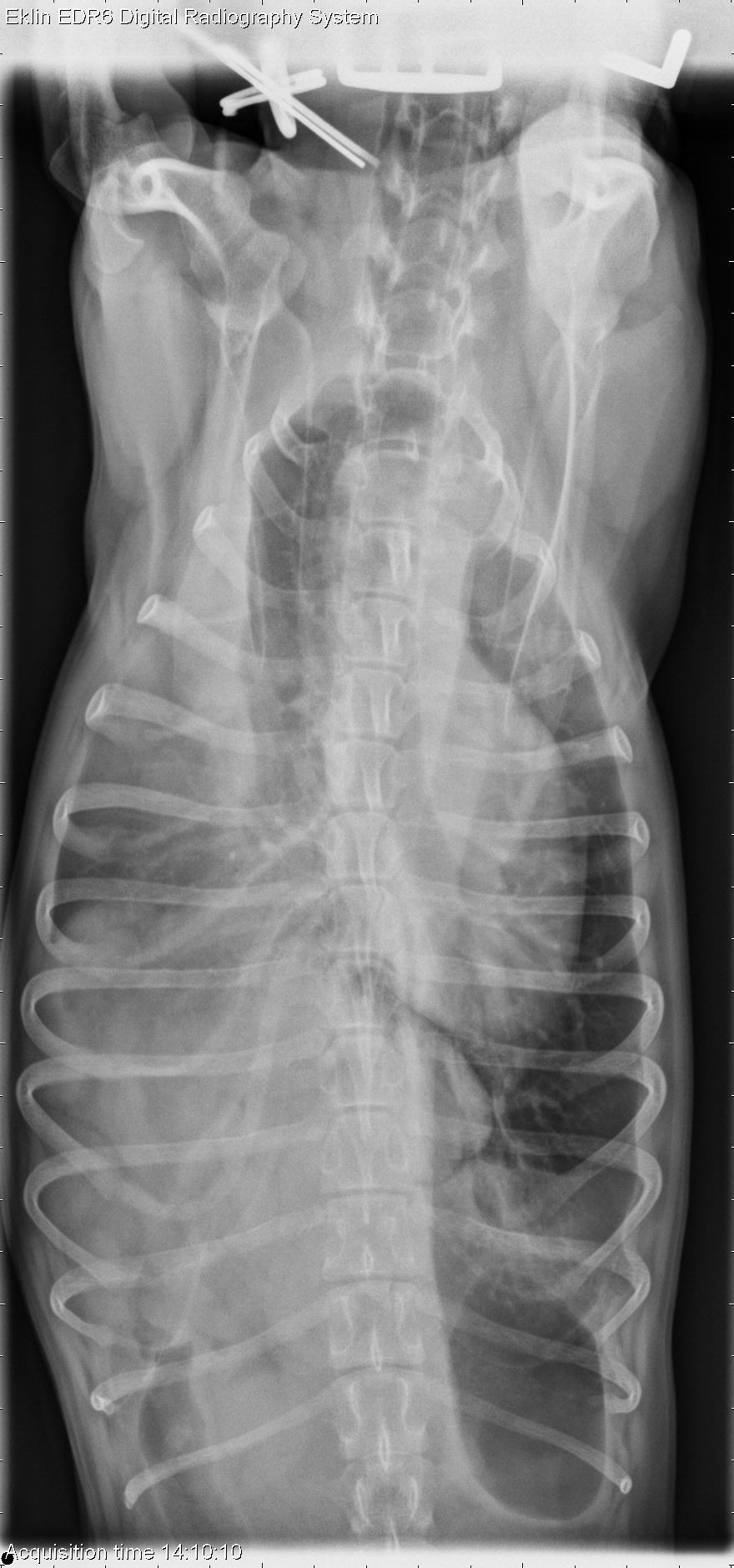
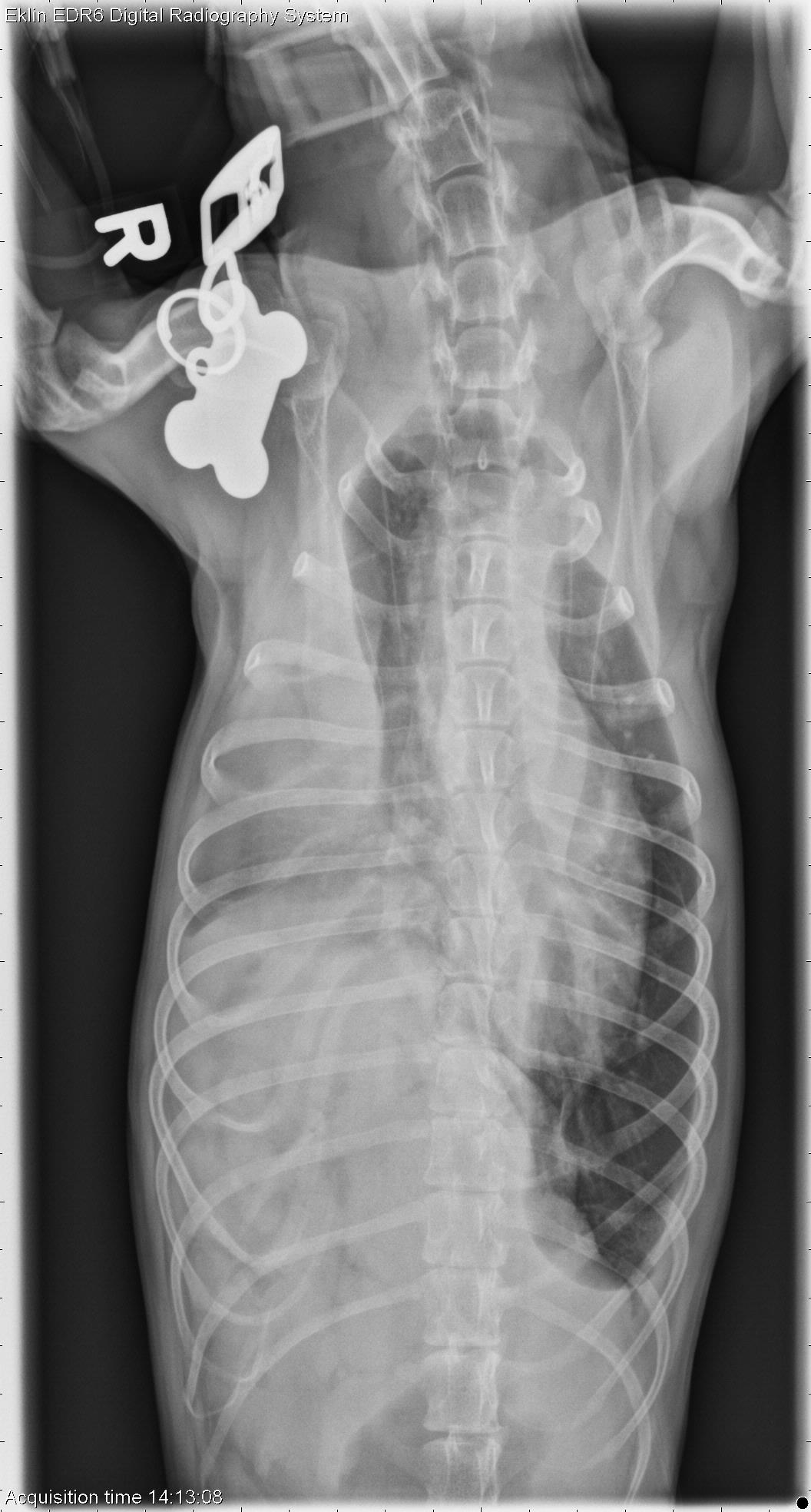
There is increased opacity in the thoracic cavity on all projections. The lung lobe margins are rounded and retracted with visible fissure lines, indicating pleural effusion. The opacities in the thoracic cavity include soft tissue and gas opacity with a granular appearance. This is mostly present on the right side, and the diaphragm is not visible on the right. The cardiac silhouette is displaced to the left by this mass effect. The left hemithorax is more normal in appearance. There is a small volume pneumothorax visible in the left caudal thorax on the ventrodorsal projection. The normal hepatic silhouette is not visible in the abdominal cavity, and the stomach is positioned more cranially. There is poor detail in the abdomen suggesting peritoneal effusion.Diagnosis
Chronic diaphragmatic hernia on the right side with secondary bicavitary effusion and small volume pneumothorax. The liver and a portion of the intestine are most likely located in the pleural space.At surgery, there were two rents in the right diaphragm with herniation of small intestine and liver.