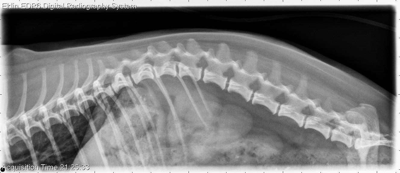
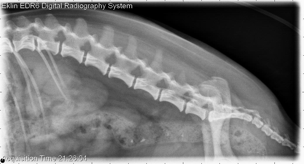
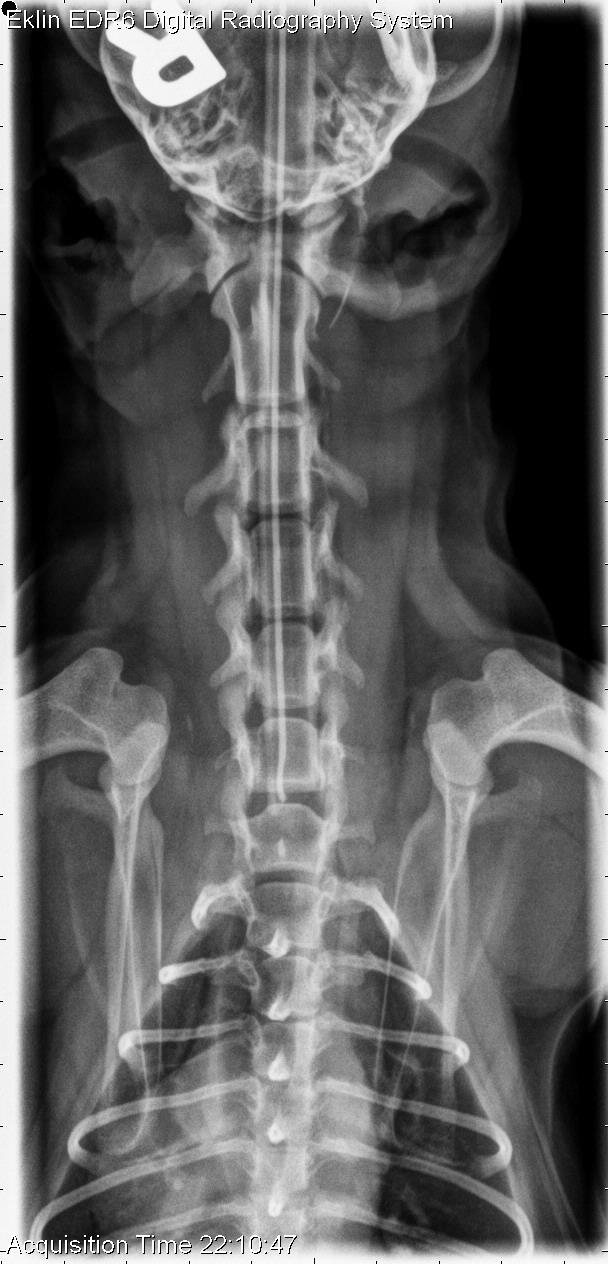
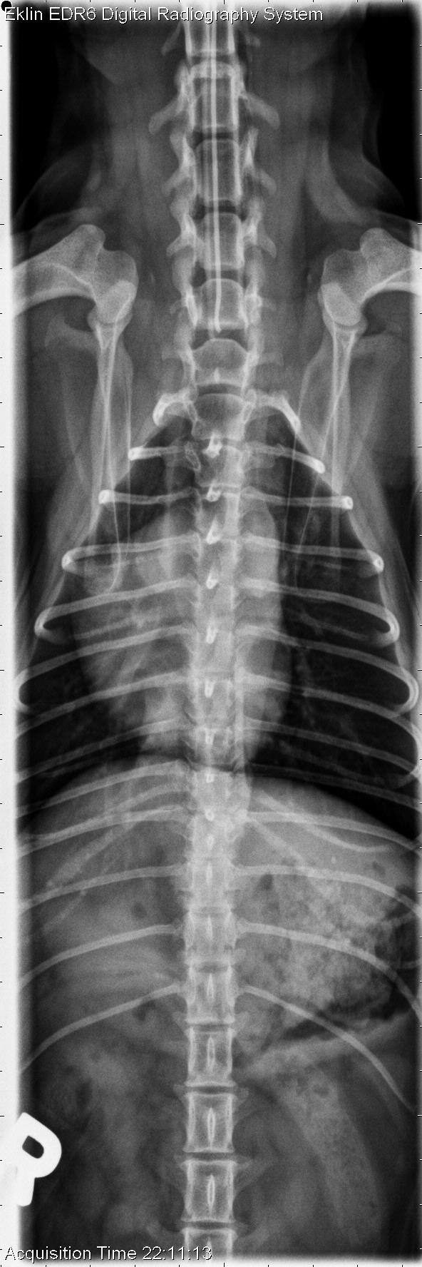
For our first case of November, we have a 4-year-old female neutered Yorkshire Terrier that was attacked by another dog. Now painful and tetraparetic. Spine radiographs are often our first imaging choice when evaluating animals for trauma. What are your findings?
Findings
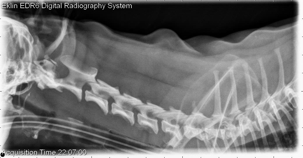
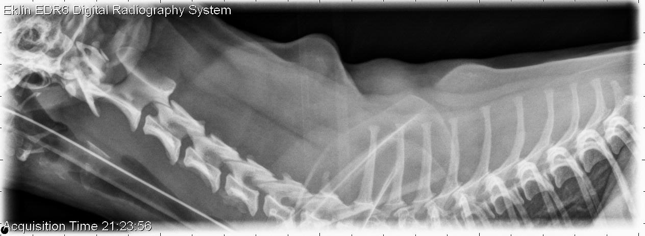
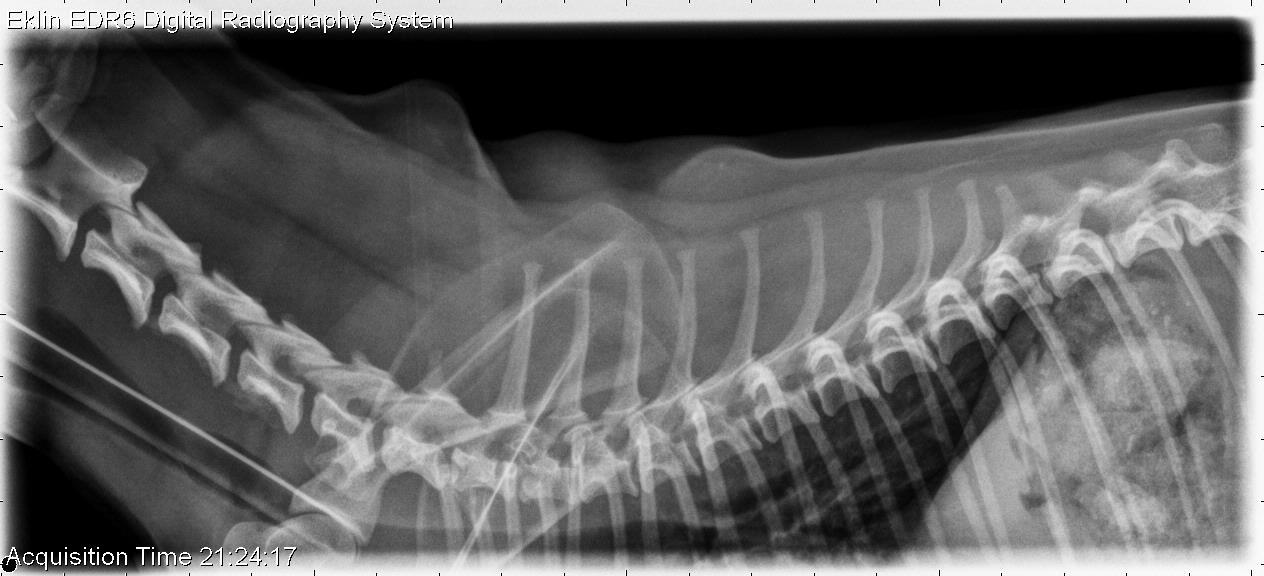
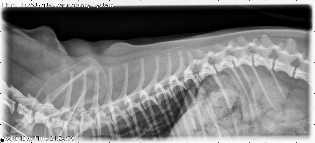
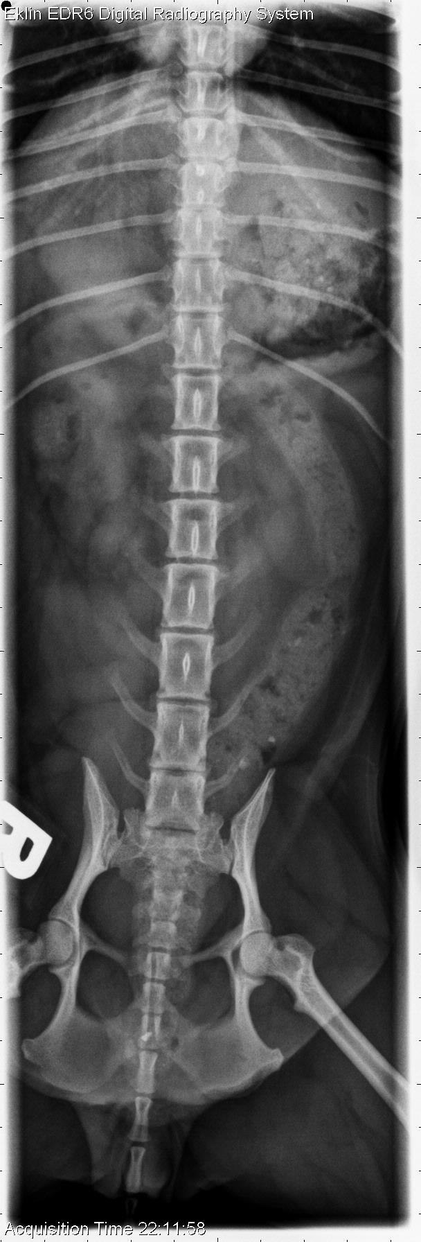
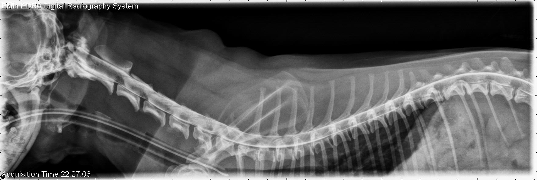
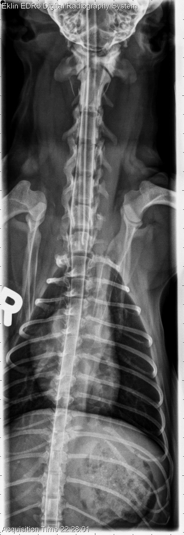
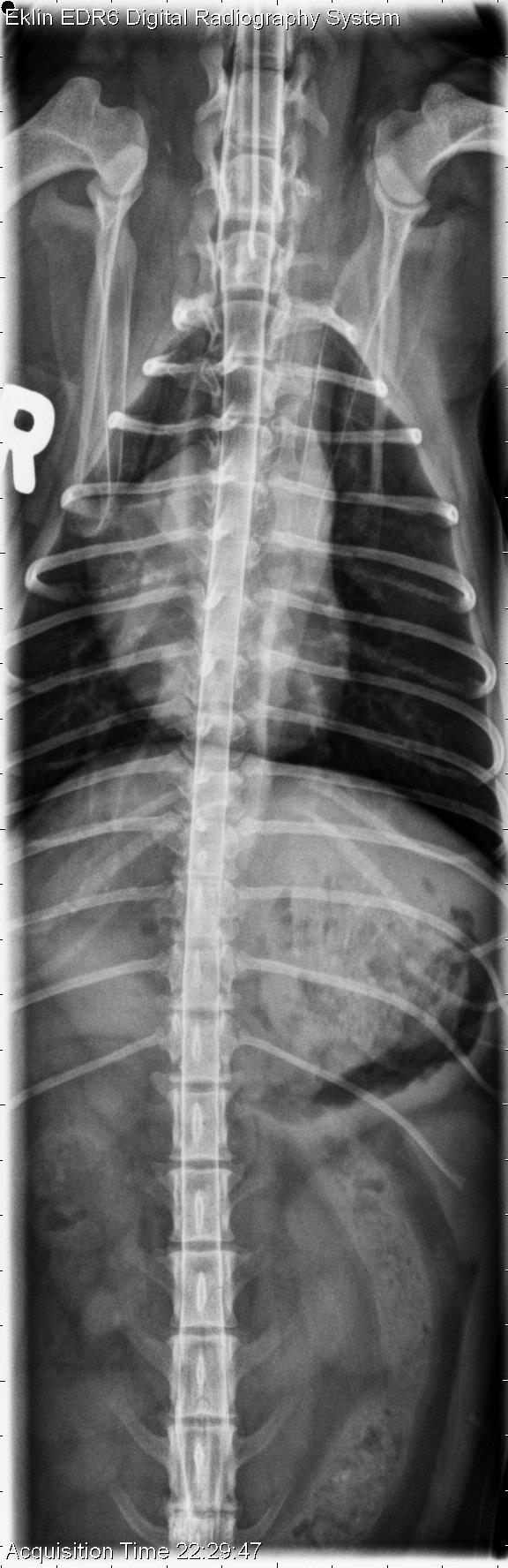
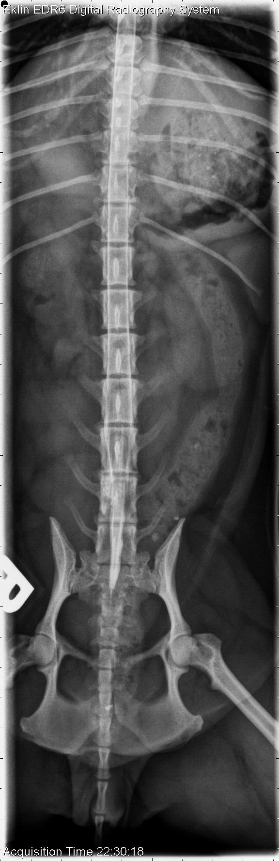
Survey spine: On the lateral images of the cervical spine, there is a bone fragment dorsal to the C1 vertebra, with a rounded opacity cranial to it. These fragments are not well seen on the ventrodorsal projections. The remainder of the spine appears normal.
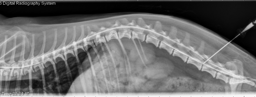
Myelogram: Contrast fills the subarachnoid space normally after an injection at L5-6. At C1-2, there is contrast leakage into the subcutaneous tissues dorsal to C1. There is splitting of the contrast column at C6-7 without attenuation of the dorsal contrast column.
Differential Diagnosis
- C1 fracture with possible luxation and dural tear
- Intervertebral disc herniation without spinal cord compression at C6-7
Diagnosis
On CT images, the fracture of C1 and dural tear were confirmed. The dog recovered the ability to walk with conservative therapy and a neck brace. On these CT images, the fractures of C1 as well as contrast in the soft tissues can be seen.