This week’s case is a 13-year-old female neutered domestic short haired cat. History of wheezing for the past 10 days. Lung lobectomy for pulmonary carcinoma two years previously. Where is the abnormality located in this case?
Findings
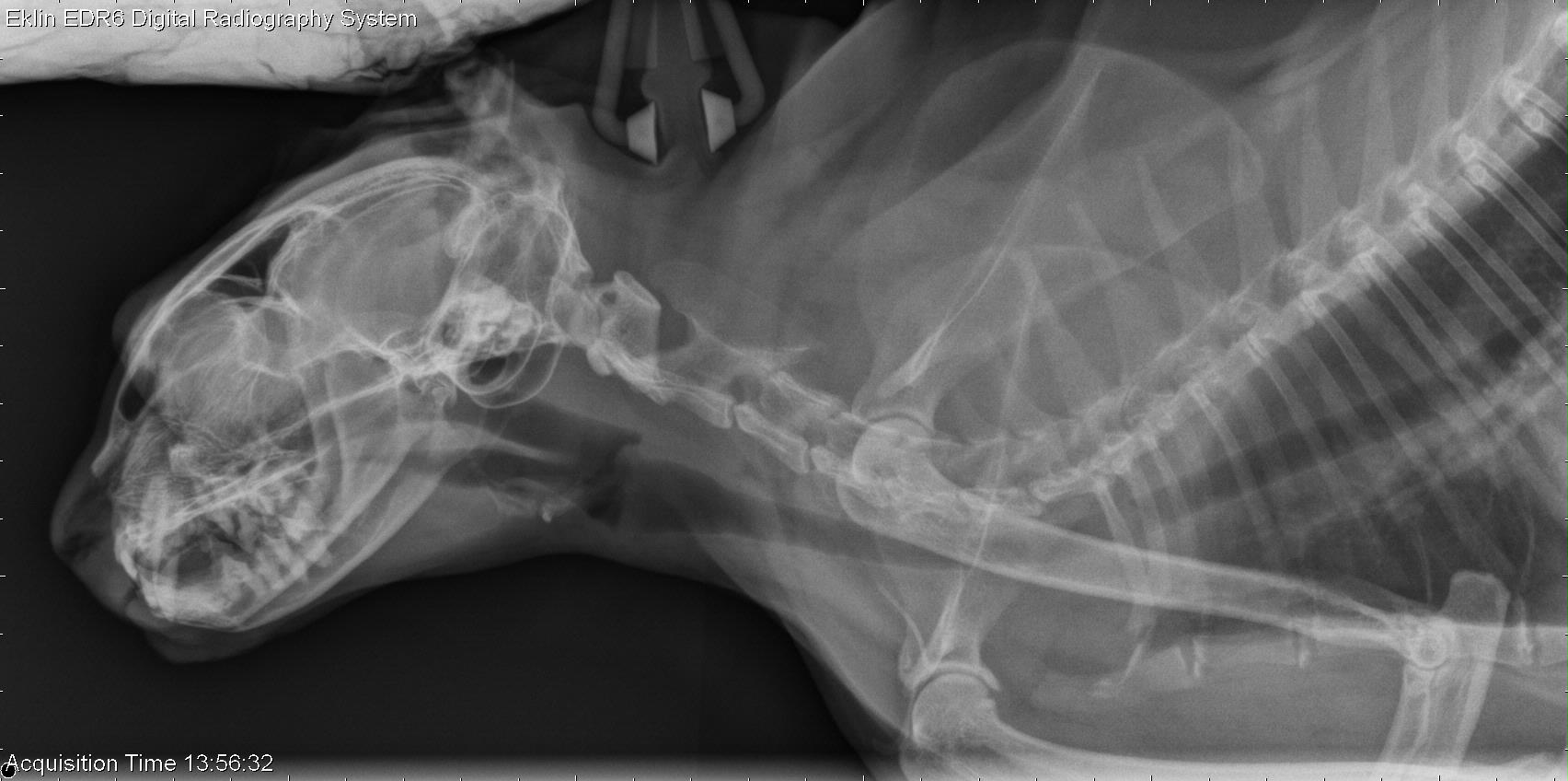
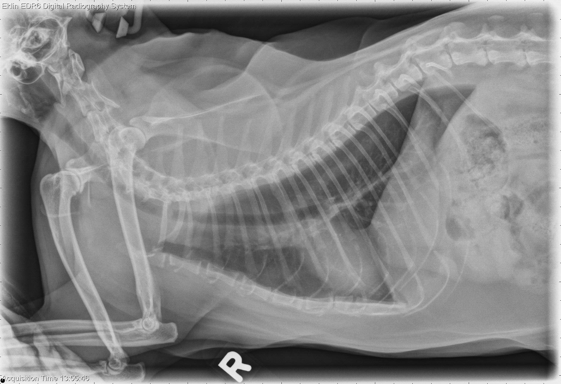
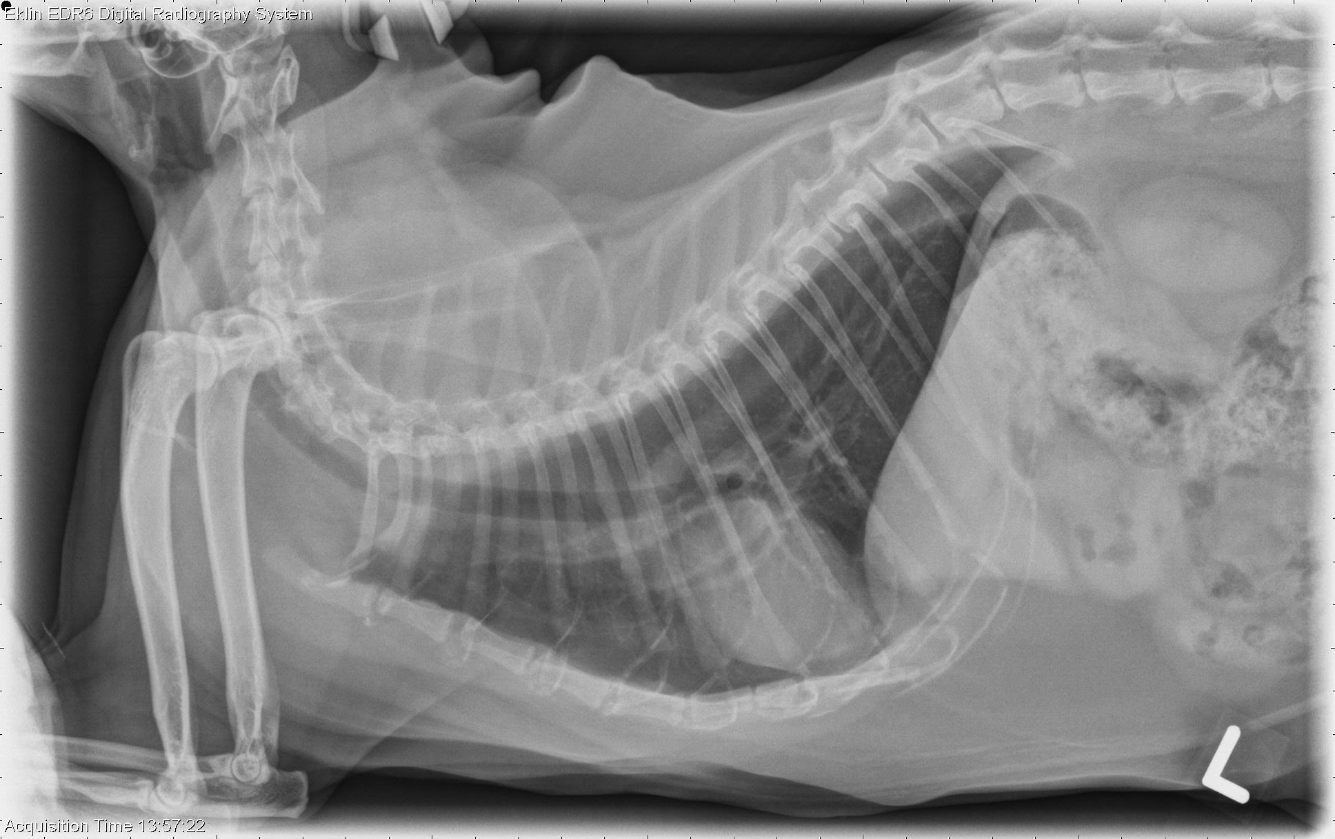
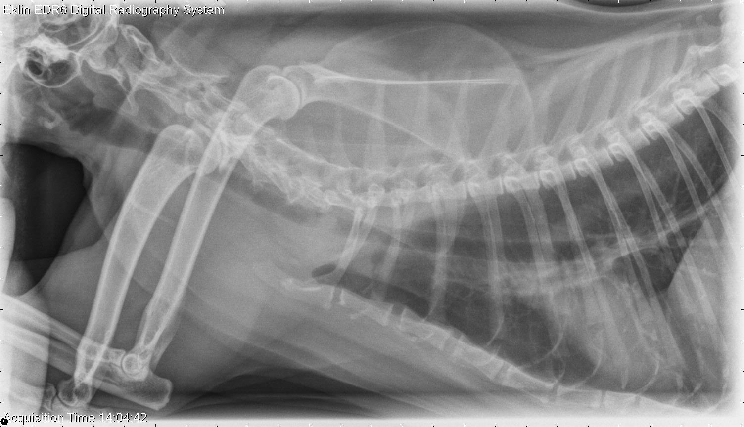
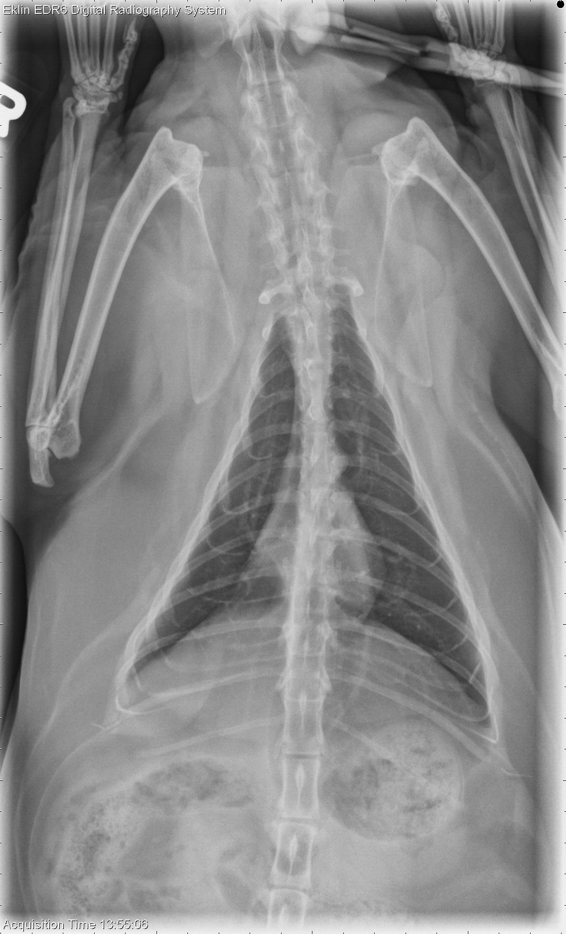
On a lateral radiograph of the neck the nasopharynx and oropharynx are distended. The soft palate and laryngeal structures appear normal. In the thorax, the cardiac silhouette is positioned in apposition to the diaphragm, likely due to the previous lung lobectomy. On both lateral projections, there is an oval soft tissue opacity outlined by gas in the tracheal lumen in the first intercostal space. This appears to occupy the entire width of the trachea, and is broad-based on the left lateral projection. The cardiovascular structures appear normal. There is mild spondylosis deformans in the caudal cervical spine, and osteophyte production in one shoulder joint. In an additional projection with the thoracic limbs pulled cranially, the mass is visible cranial to the first rib within the tracheal lumen.Differential Diagnosis
- Intraluminal tracheal mass (neoplasia, granuloma) likely causing the wheezing
- Distended pharynx secondary to upper airway obstruction
- Incidental shoulder osteoarthrosis
Diagnosis
- Metastatic pulmonary carcinoma
- The previously removed pulmonary carcinoma had invaded the right caudal lobar bronchus and was incompletely exised