This week’s case is a 13-year-old male neutered West Highland White terrier. The owner found blood on the carpet this morning. Crackles ausculted in right thorax. Seen previously for polypoid gastric mass. Where do you think the mass is originating from? What are the differential diagnoses? Feel free to post questions and comments below.
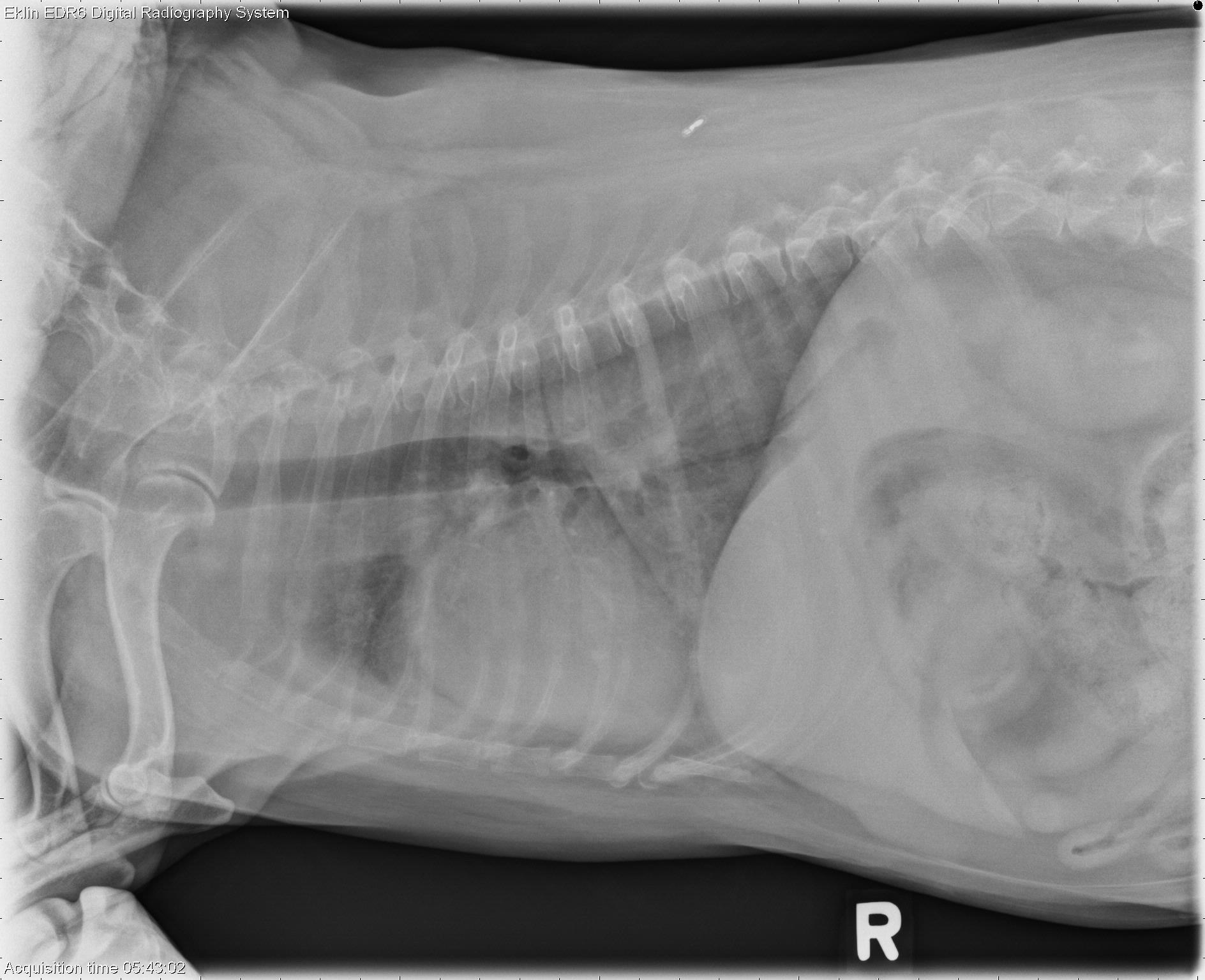
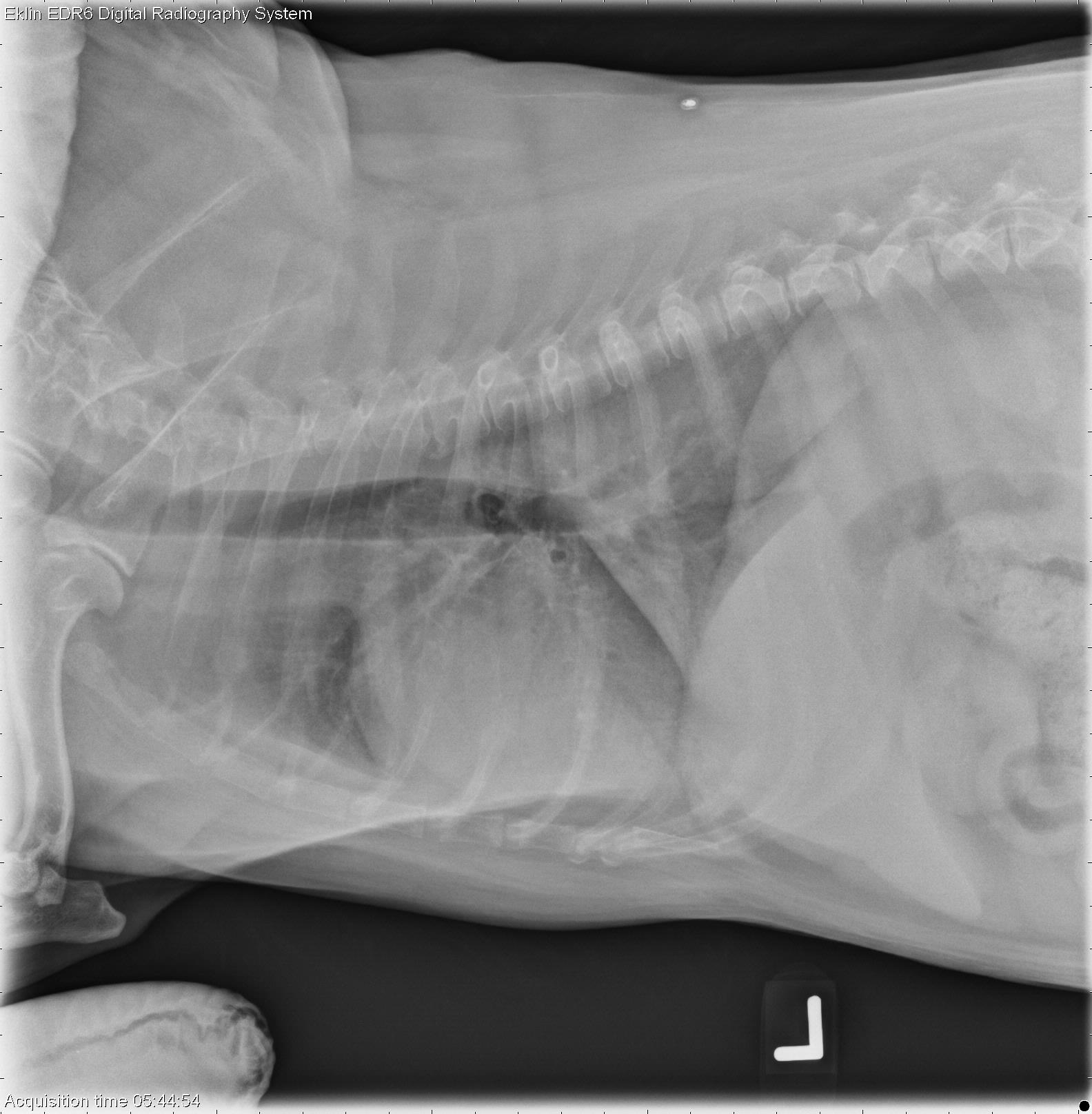
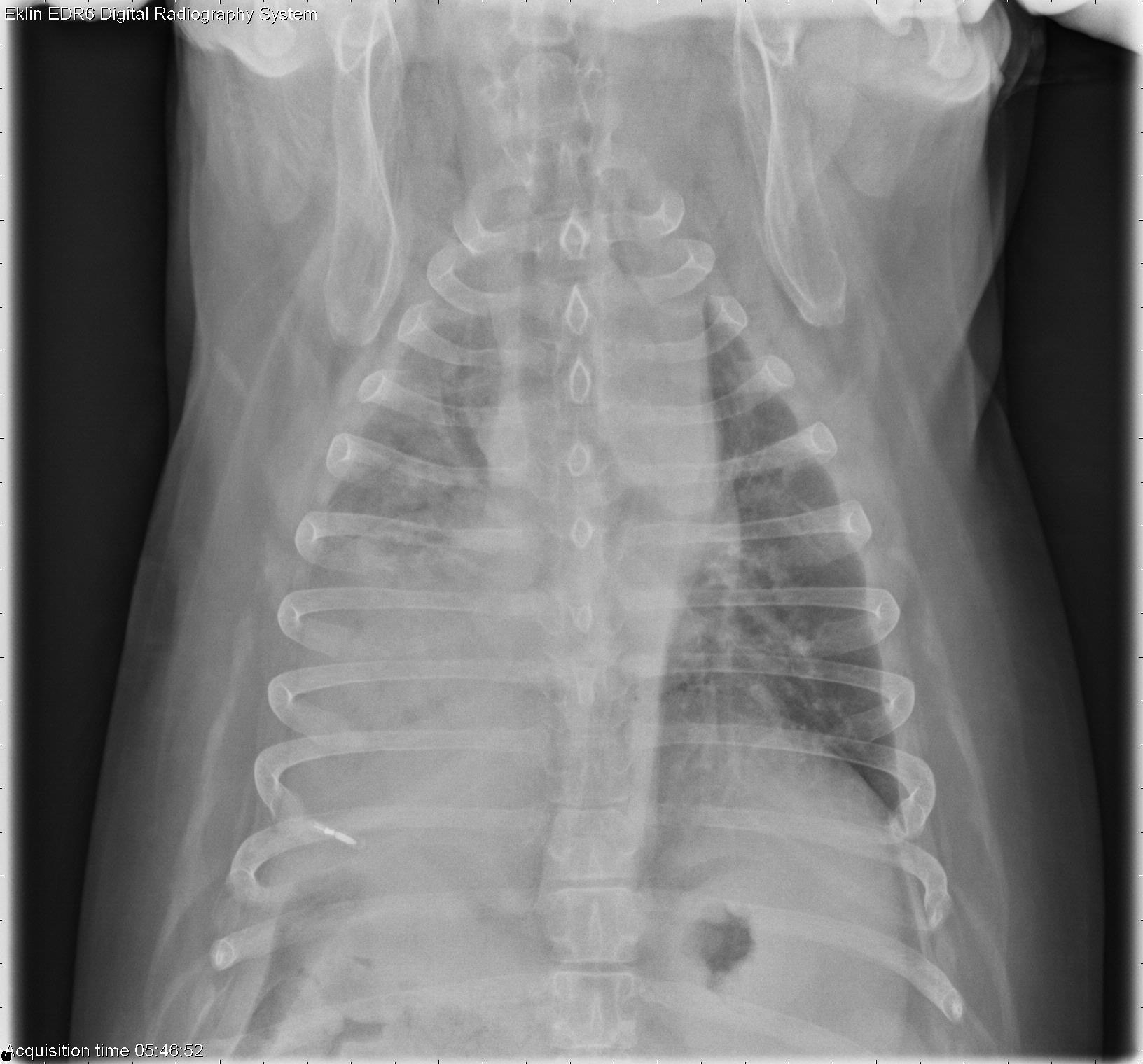
Findings
There is an alveolar pattern occupying the entire right caudal lung lobe, visible on all three projections of the thorax. The broncus to the right caudal lung lobe is poorly visualized. There is additional interstitial opacity in the right middle and right cranial lung lobes. The left lung appears normal. The cranial mediastinum is widened; however, this is likely a breed variation. The cardiac silhouette appears normal in size and shape. The pulmonary vasculature is normal. In the portion of the abdomen included, the pylorus is soft tissue opacity on both lateral projections.
Differential Diagnosis
- Right caudal lobar consolidation – primary pulmonary neoplasia, hemorrhage
- Cranial lobe interstitial pattern – hemorrhage, atelectasis
- Pyloric soft tissue opacity – likely known gastric polypoid mass (polyp, leiomyoma)
Diagnosis
- Pulmonary squamous cell carcinoma
- Gastric leiomyoma