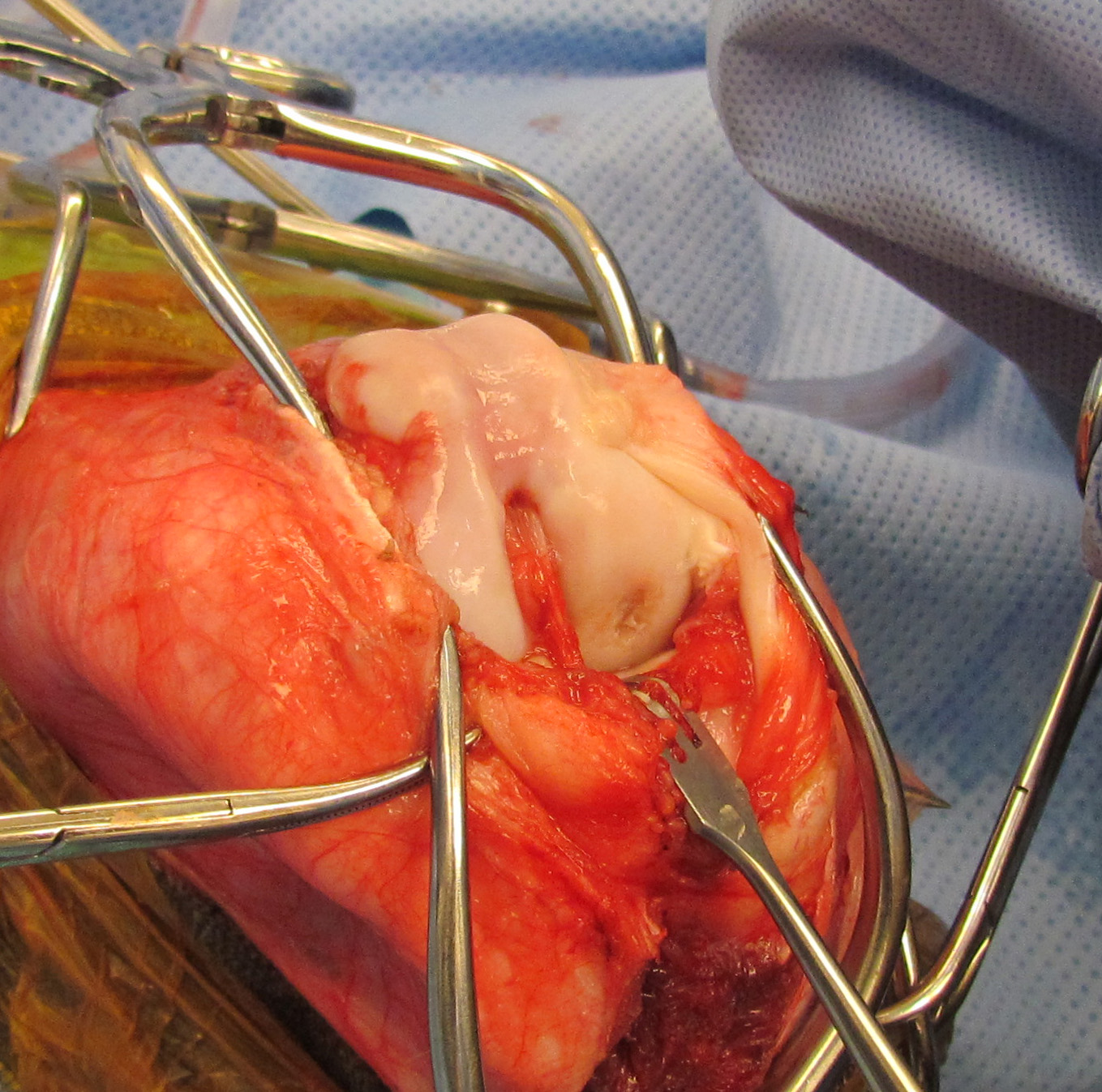
Today’s case is a 9-month-old male Rhodesian Ridgeback who has been lame on the left pelvic limb for three weeks. Take a look and post your interpretation.
Findings
Left stifle: There is lateral luxation of the patella, and the trochlear grooves of the femur appear shallow. There is increased soft tissue effusion within the joint. On both projections, there is a radiolucent defect in the lateral condyle of the femur involving subchondral bone. Small, rounded mineral opacities are visible in the caudal aspect of the joint. There are linear, horizontal lines visible in the medulla of the femur.
Right stifle: There is moderately increased soft tissue opacity within the joint. The lateral femoral condyleis also flattened with a small radiolucent defect, less severe than the left. There are linear, horizontal lines visible in the medulla of the femur.
Differential Diagnosis
- Osteochondrosis dissecans lesion in the left lateral femoral condyle with fragments in the joint
- Osteochondrosis in the right stifle
- Bilateral stifle effusion likely due to OCD
- Left lateral patellar luxation
- Horizontal lines represent “growth arrest” lines and are incidental
Diagnosis
The left stifle was surgically explored. A large defect in the lateral condyle was debrided and treated with an OATS (autogenous osteochondral grafting) procedure. The luxating patella was repaired with a 4-in-1 procedure.