This week’s case is a 14-year-old female neutered Golden Retriever with progressive paresis of both hind limbs.
Findings
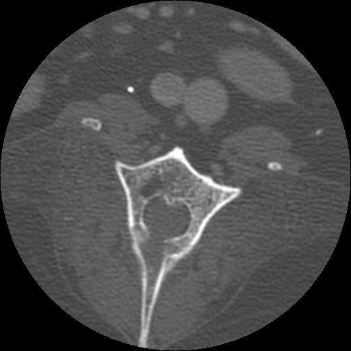
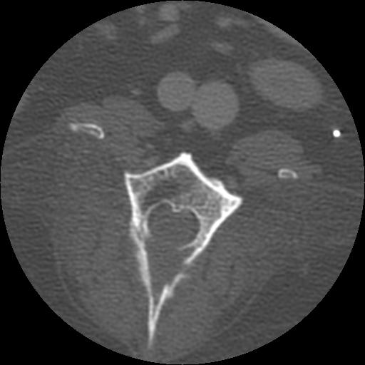
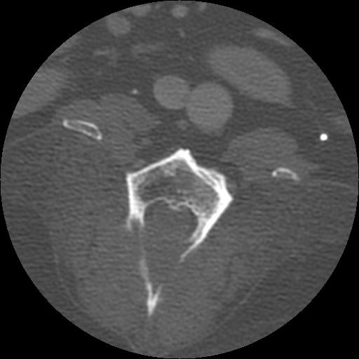
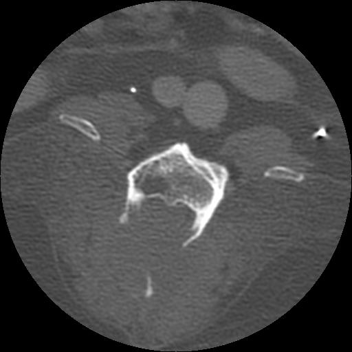
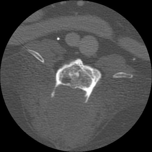
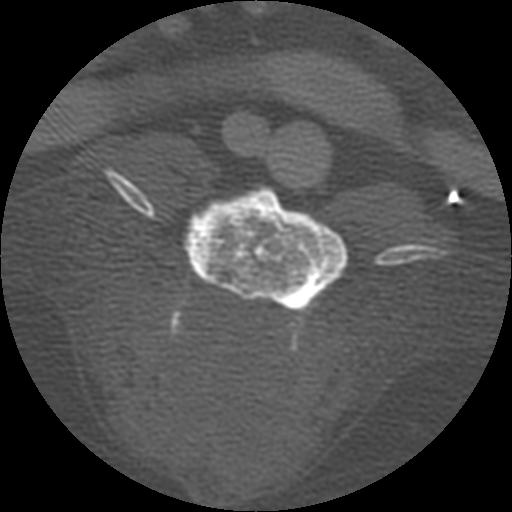
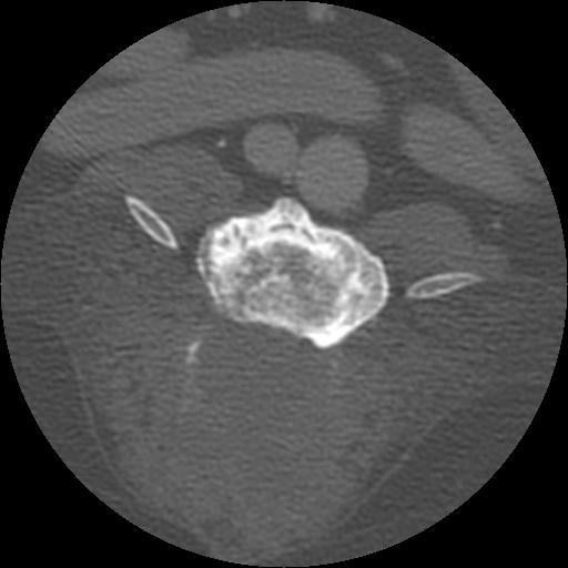
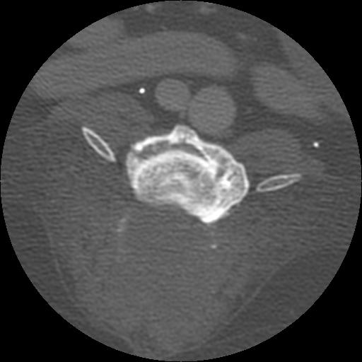
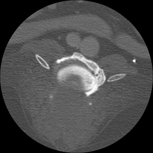
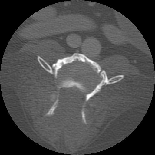
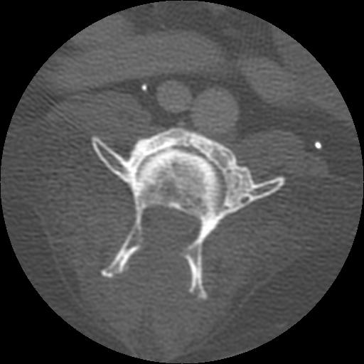
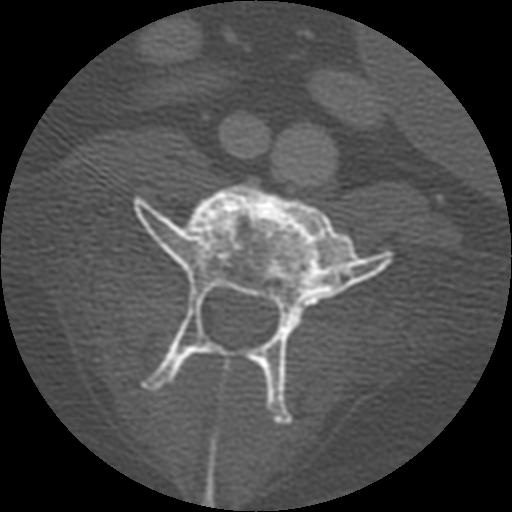
On the lateral radiograph, a large osteolytic site is present at the caudal aspect of the lamina of L4. This results in loss of the caudal half of the lamina and the articular facets of L4 with loss of the pedicles evident on the ventrodorsal projection. There is dural ossification in the cord. There is extensive ventral spondylosis throughout the lumbar vertebral column. No other sites of osteolysis are identified.On the 1 mm, post contrast CT images, there is extensive osteolysis in L3 and L4 involving the vertebral bodies, laminae and dorsal spinous processes. There is a non contrast-enhancing soft tissue mass dorsal to the vertebrae.
Differential Diagnosis
- soft tissue sarcoma
- round cell neoplasia
- other soft tissue tumor invading bone