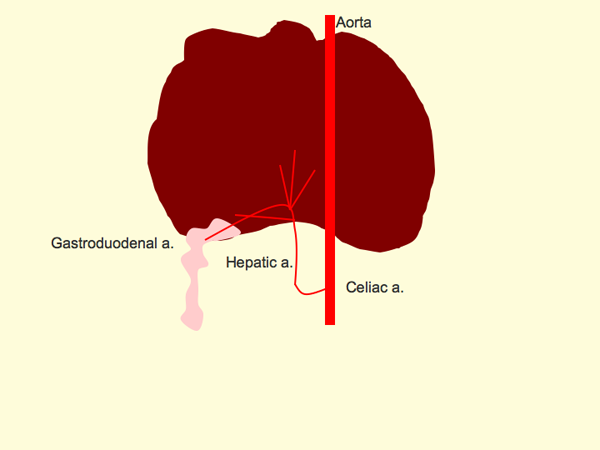
For those of you doing ultrasound in practice, there are some fundamental facts about the vascular anatomy of the liver that are useful to know. The liver has a dual blood supply, with 75% coming from the portal vein, and 25% from the hepatic artery. The portal vein delivers nutrient-rich and oxygen-poor blood from the intestines and splanchnic organs, and the hepatic artery supplies oxygenated blood. Both of these blood supplies mix in the hepatic sinusoids to provide the liver with the oxygen, nutrients and growth factors that it needs.
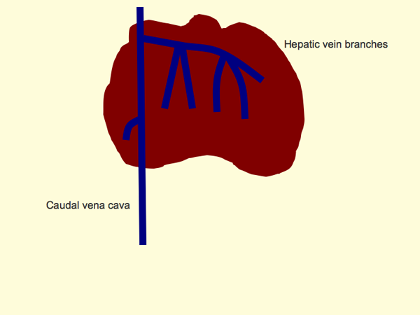
For drainage, there is a single system of hepatic veins that connect to the caudal vena cava. There are some small hepatic veins on the right in the dorsal part of the liver, and a large system of veins draining the right medial, quadrate and left liver lobes.

So there are two types of vessels entering the liver at the hilus, and one type leaving the liver close to the diagphragm. Within the parenchyma, these vessels tend to interlace, like the fingers of your hands if you clasp them in front of you. How do you tell them apart?
The hepatic arteries are generally not visible except for very close to the hilus. You may pick them up with Doppler ultrasound. The arteries run parallel to the portal veins on either side, which can help to identify their location.
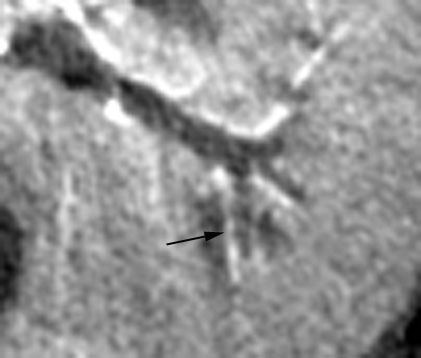
This CT image shows the portal veins (dark grey) parallelled by hepatic arteries (arrow). So the two types of vessels entering the liver will have the same direction of flow with Doppler; towards the center and periphery of the liver.
The hepatic veins drain the liver, and originate as small branches in the periphery, coalescing to larger vessels near the caudal vena cava. The left hepatic vein is the largest, with several large branches. In the mid portion of the liver, the peripheral hepatic veins and portal veins interlace. The key to telling them apart is the wall characteristics.
The portal veins have hyperechoic walls because of fat and fibrous tissue. The walls are generally visible and distinct, even if the vessel is a smaller one. In contrast, the hepatic veins have thinner walls with less of a hyperechoic signal. In addition, where the two types of veins interlace, the Doppler signal will be running in opposite directions.
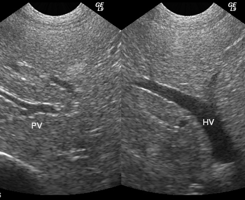
In this image, the much larger hepatic vein has less distinct walls than the small portal vein. At the hilus, the portal vein is also surrounded by fat, which can help to identify where it enters the liver.
Vascular anatomy of the liver is important in several situations. The most potentially frustrating is the search for a portosystemic shunt. It really helps to know the normal anatomy before looking for abnormal vessels. It also comes into play when looking for changes in echogenicity of the liver. In a hyperechoic liver, the portal vein walls are not visible as far peripherally as usual. In a hypoechoic liver, they are visible right to the periphery. The hepatic vessels are also useful as landmarks to tell approximately which liver lobe you are looking at. Take a look for them next time you scan.
Recent Comments