This week’s case is a 3-year-old female neutered Yorkshire Terrier with 1-week history of lethargy, dyspnea, and decreased appetite. The second set of radiographs was taken two weeks post-treatment. Post your interpretations and differentials in the comments section!
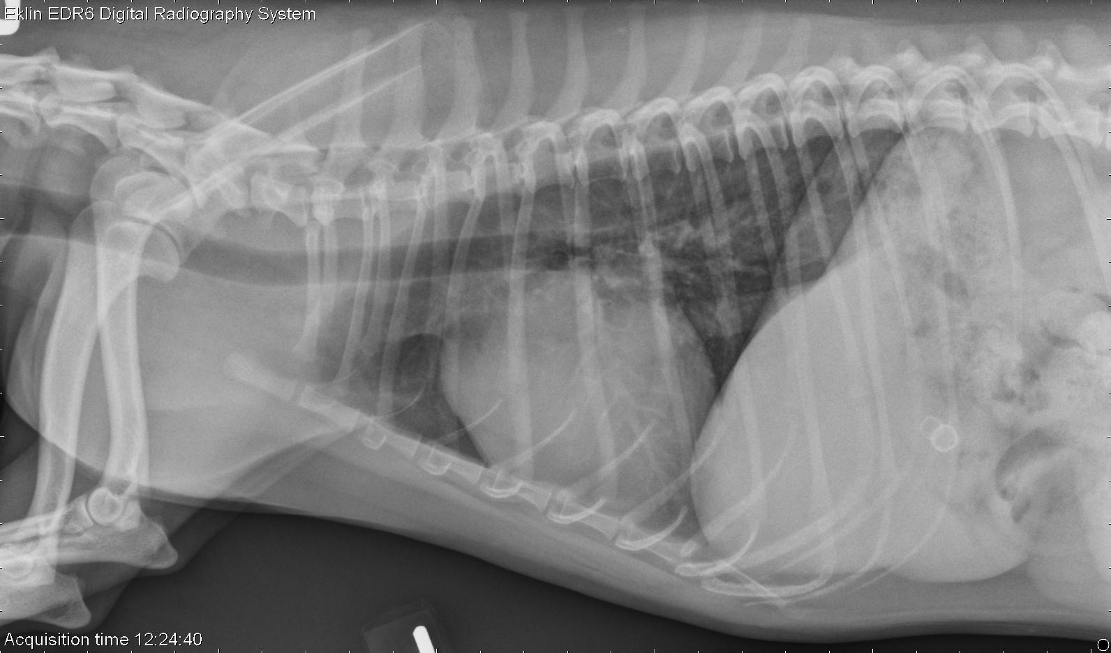
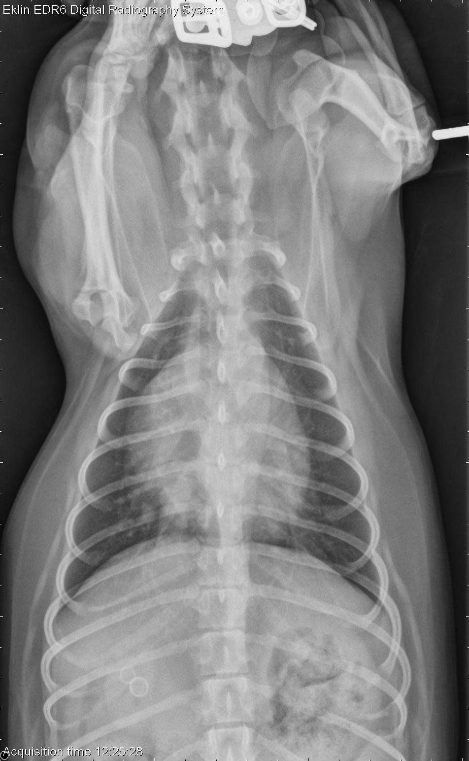
Findings
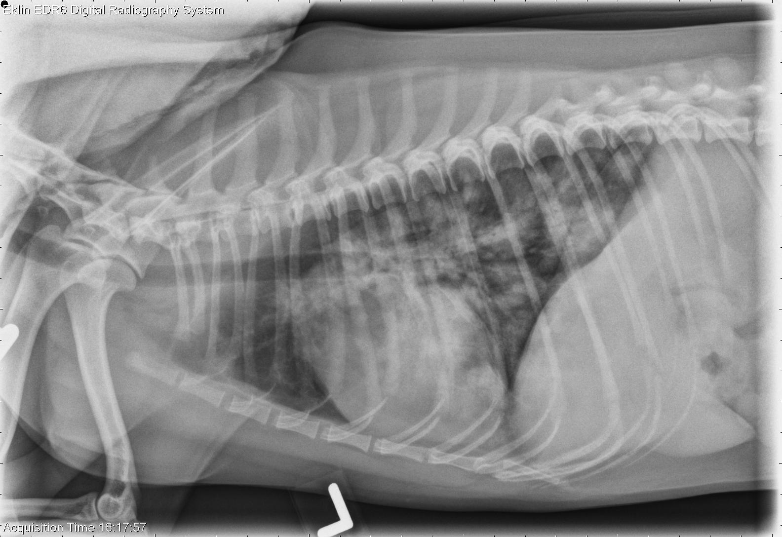
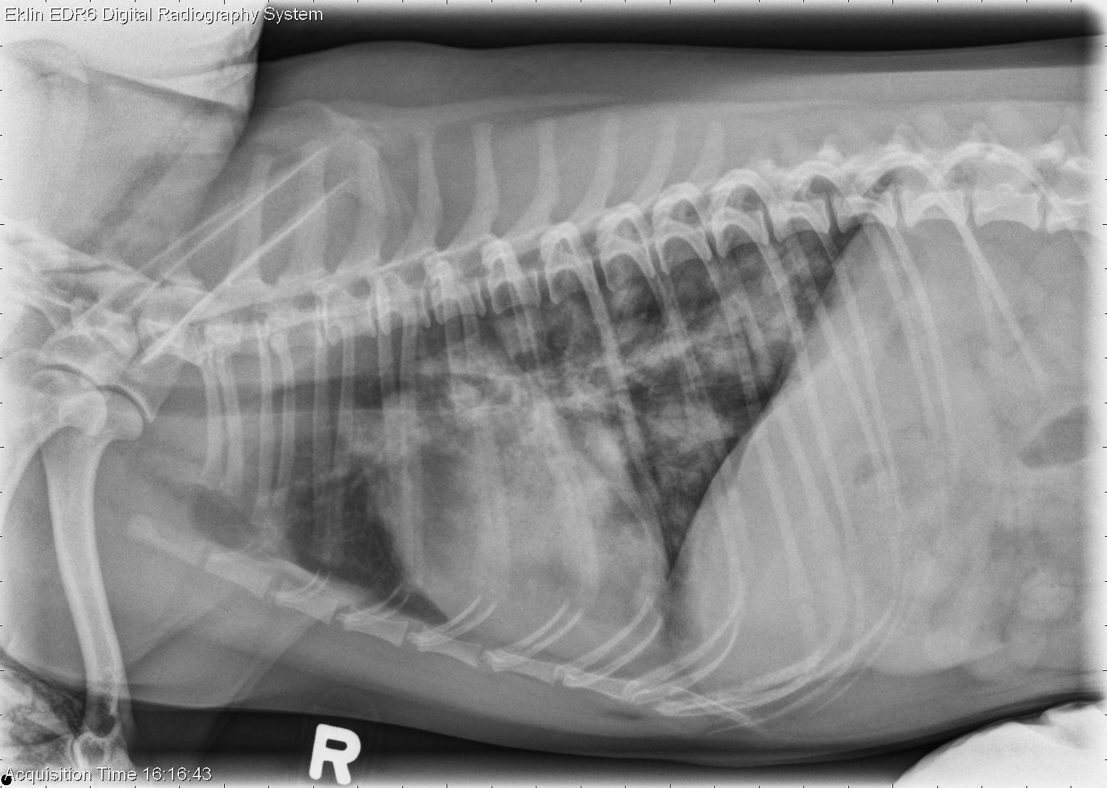
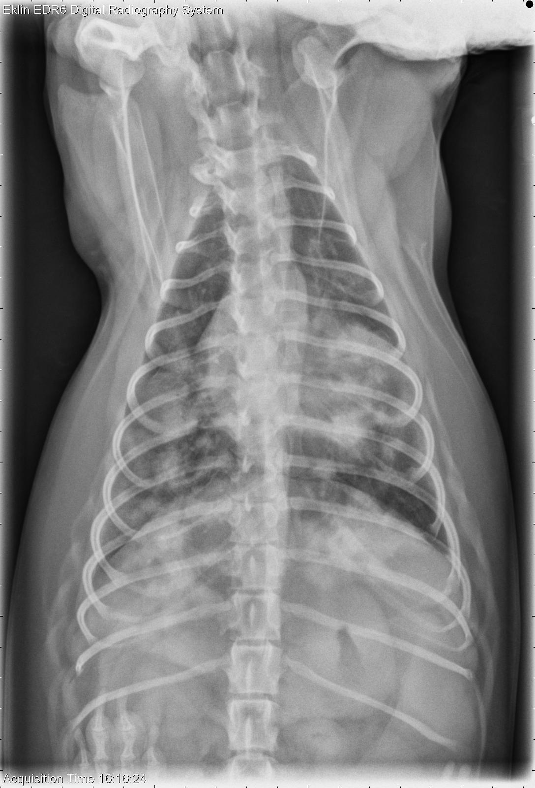
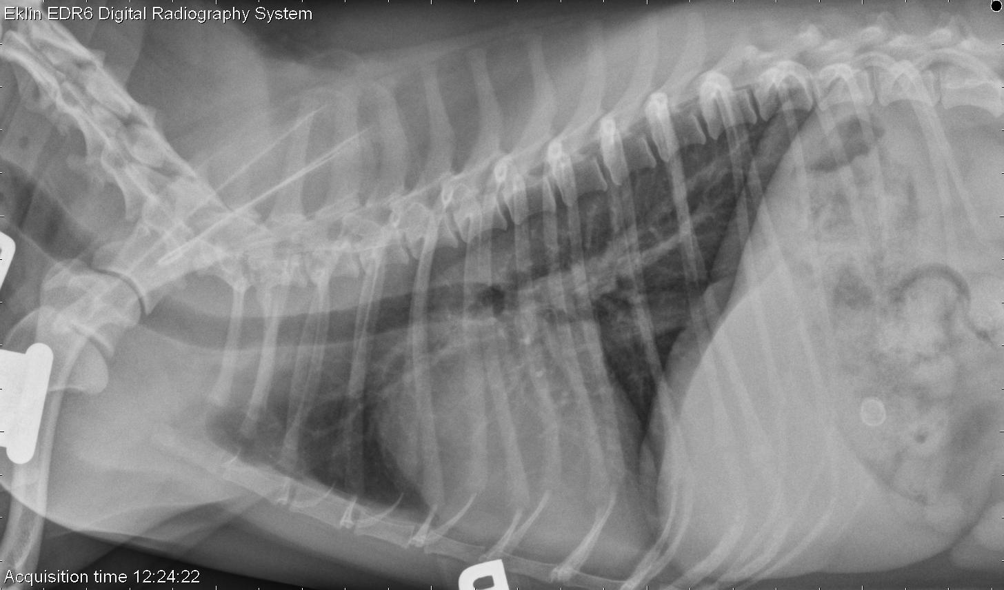
There is a patchy alveolar pattern with ill-defined soft tissue opacities throughout the lungs. The pattern is most prominent in the caudodorsal lung. There is no evidence of lymphadenopathy, and the cardiovascular structures are normal.The second series was taken two weeks later after treatment. The pulmonary pattern has resolved.
Differential Diagnosis
- embolic pneumonia
- parasitic pneumonia
- PTE
- hemorrhage
- fungal pneumonia
- other infectious/inflammatory disease
Diagnosis
Steroid responsive inflammatory lung diseaseDiscussion
Lung aspirates were taken and showed moderate to high numbers of nucleated cells, most of which were neutrophils with some lymphocytes. The pathologic diagnosis was moderate to marked mixed, primarily suppurative, inflammation with mild epithelial hyperplasia. An underlying infectious etiology was felt to be most likely, though no organisms were found on an extensive search. Fungal titres were negative.The dog was treated with antibiotics and steroids, and improved greatly within two weeks. She is being weaned off steroids and monitored closely.