• Home
  • About
  • Articles
  • Books
  • Contact

Veterinary Radiology

Teaching and learning about veterinary diagnostic imaging.

  • Modality
    • Radiographs
    • Ultrasound
    • CT/MRI
  • Region
    • Thorax
    • Abdomen
    • Musculoskeletal
    • Neurologic
  • Species
    • Canine
    • Feline
    • Equine
    • Exotic

Biliary obstruction

November 13, 2007 By Allison Zwingenberger

Extrahepatic biliary obstruction is a reasonably common clinical scenario in older cats. Chronic cholangiohepatitis, pancreatitis and neoplasia can all cause bile duct obstruction with jaundice and high serum bilirubin. The bile duct can undergo a chronic partial obstruction from a mass or inflammatory tissue, or an acute obstruction from a calculus.

In complete biliary obstruction, the gallbladder is enlarged. The cystic duct and common bile duct become dilated and tortuous (image 1). The normal common bile duct should measure less than 4 mm in diameter. Trace it to the duodenal papilla to check for calculi and masses (image 2). The papilla should also be less than 4 mm. The one pictured here measured 3.6 mm.

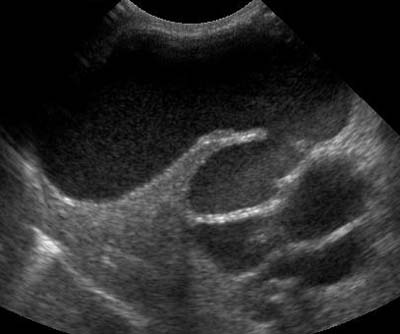
Once the complete obstruction has been present for a week, the intrahepatic bile ducts also dilate. In image 3, the intrahepatic bile duct is the anechoic structure next to the gall bladder. When color Doppler is applied, there is no flow, though it is similar in size to portal and hepatic veins. The inner surface of the wall is also slightly irregular which may represent inflammatory tissue or calculus.

Nyland TG, Gillett NA. Sonographic evaluation of experimental bile duct ligation in the dog. Veterinary Radiology 1982;23:252-260.
Biliary obstructionDuodenal papillaIntrahepatic bile duct

Filed Under: Ultrasound Tuesday

  • Bloglovin
  • Facebook
  • RSS
  • Twitter
AtlasCover787x1024 amazon-availablenow

Veterinary Radiology News

Sign up for alerts about new cases and newsletters.

We will respect your privacy.

Archives

Recent Comments

Tags

abscess adenocarcinoma bronchiectasis carcinoma cardiomyopathy coccidioidomycosis discospondylitis elbow dysplasia eosinophilic bronchopneumopathy erosive polyarthritis feline infectious peritonitis FIP foreign body fracture gastric dilation gastric foreign body gastrointestinal GDV heart failure hemangiosarcoma hiatal hernia histiocytic sarcoma hypertrophic cardiomyopathy intestinal foreign body linear foreign body lymphoma megaesophagus osteochondrosis osteomyelitis osteosarcoma patent ductus arteriosus PDA pericardial effusion pneumonia pneumothorax polyarthritis PPDH pulmonary pulmonary abscess pulmonary adenocarcinoma pulmonary carcinoma sarcoma thymoma tracheal collapse vascular ring anomaly

Copyright © 2025 · Metro Pro Theme On Genesis Framework · WordPress · Log in