• Home
  • About
  • Articles
  • Books
  • Contact

Veterinary Radiology

Teaching and learning about veterinary diagnostic imaging.

  • Modality
    • Radiographs
    • Ultrasound
    • CT/MRI
  • Region
    • Thorax
    • Abdomen
    • Musculoskeletal
    • Neurologic
  • Species
    • Canine
    • Feline
    • Equine
    • Exotic

Acute renal infarcts

October 30, 2007 By Allison Zwingenberger

Renal infarcts are often incidental findings in animals with chronic renal disease. Infarcts are caused by thrombi that occlude a blood vessel in the kidney. Most of the time, they are chronic and mainly made up of fibrous tissue. The classic appearance is of a hyperechoic, wedge shaped area with the wide part of the triangle at the kidney capsule. The capsule is also usually concave at the site because of fibrosis and loss of normal renal parenchyma.

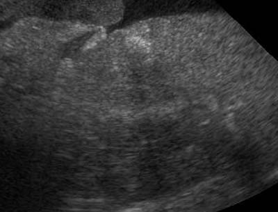
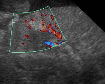
Acute renal infarcts have the same etiology, but we are seeing them within a few days after they occur. What do they look like? They are more subtly hyperechoic than the chronic infarct. In image 1, the infarct is visible as an indistinct, hyperechoic wedge in the cortex of the kidney. The medulla is out of plane so most of what is visible is cortex. The second image is a color Doppler display of the blood flow in the renal vessels. You can see the signal disruption and partial void in the hyperechoic are that represents the infarct. This dog had a systemic neoplasia with infarcts affecting the kidneys and other organs. Acute renal infarcts don’t alter the shape of the renal cortex.

Renal infarctRenal infarct

Filed Under: Ultrasound Tuesday

  • Bloglovin
  • Facebook
  • RSS
  • Twitter
AtlasCover787x1024 amazon-availablenow

Veterinary Radiology News

Sign up for alerts about new cases and newsletters.

We will respect your privacy.

Archives

Recent Comments

Tags

abscess adenocarcinoma bronchiectasis carcinoma cardiomyopathy coccidioidomycosis discospondylitis elbow dysplasia eosinophilic bronchopneumopathy erosive polyarthritis feline infectious peritonitis FIP foreign body fracture gastric dilation gastric foreign body gastrointestinal GDV heart failure hemangiosarcoma hiatal hernia histiocytic sarcoma hypertrophic cardiomyopathy intestinal foreign body linear foreign body lymphoma megaesophagus osteochondrosis osteomyelitis osteosarcoma patent ductus arteriosus PDA pericardial effusion pneumonia pneumothorax polyarthritis PPDH pulmonary pulmonary abscess pulmonary adenocarcinoma pulmonary carcinoma sarcoma thymoma tracheal collapse vascular ring anomaly

Copyright © 2025 · Metro Pro Theme On Genesis Framework · WordPress · Log in